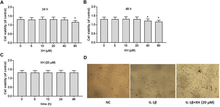
FIGURE 2.
Cell viability of XH on rat chondrocytes. CCK8 assays were conducted to determine the effects of XH (0, 5, 10, 20, 40, and 80 μM) in 24 h (A) and 48 h (B), respectively. (C) The effects of XH (20 μM) on cell viability in 48 h. (D) The cell morphology was observed after administration with IL-1β (10 ng/ml) with or without XH (20 μM) in 48 h. All experiments were performed in triplicate and data are presented as the mean ± standard deviation. * p < 0.05 and **p < 0.01.

