Figure S3.
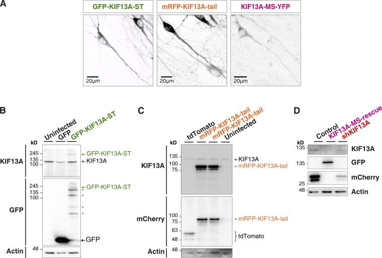
Expression of the recombinant proteins GFP-KIF13A-ST, mRFP-KIF13A-tail, and KIF13A-MS-YFP. (A) Representative confocal images of GFP-KIF13A-ST (green), mRFP-KIF13A-tail (orange), and KIF13A-MS-YFP (pink) expressed in organotypic hippocampal slices. (B) Western blot analysis of GFP-KIF13A-ST expression with antibodies against GFP and KIF13A. Infection with a GFP virus is used as control. Actin is used as loading control. (C) Similar Western blot analysis with slices expressing mRFP-KIF13A-tail or tdTomato, as control. (D) Western blot analysis of dissociated hippocampal neurons showing the reduction on the endogenous KIF13A protein levels (shKIF13A and KIF13A-MS-rescue conditions; KIF13A antibody) together with the expression of the recombinant and shKIF13A-resistant KIF13A-MS-YFP protein (KIF13A-MS-rescue condition; GFP antibody). Recombinant KIF13A-MS-YFP is not recognized by the KIF13A antibody, as the epitope is located in the absent globular tail domain. Both control (lacking the shRNA sequence) and shKIF13A lentiviruses express mCherry. Actin is used as loading control.

