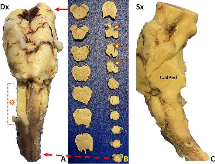
Fig. 1.
Macroscopic brainstem appearance in COVID-19-patients. A Anterior surface after major arterial vessel removal: no evidence of pathological changes. Dx: right side. Asterisk: right medulla oblongata (bulb) area removed for tissue sampling. B Transverse brainstem sections. No evidence of gross pathological changes. The solid arrow (top) indicates the section at cerebral peduncle and the substantia nigra levels. The asterisks correspond to the sampled area in image A. The dashed arrow (bottom) corresponds to the most caudal medulla oblongata section. C Left brainstem (dorsal face on the right). C.alPed: cerebellar stalk

