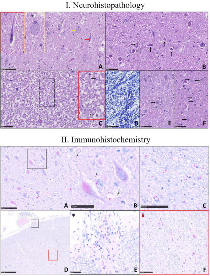
Fig. 2.
Microscopic brainstem examination. I. Images are obtained by Nanozoomer Hamamatsu s360 and magnification is represented as scale bare. Histopathological features of nervous tissue damage in the pons and medulla oblongata (MO; scale bare: 50 µm). A–D and F are from COVID-19 patients. A Pons, V motor nucleus. Different types of neuronal damage: neurons with condensed chromatin and shrunken cell body (black arrow), and neuronal chromatolysis (yellow arrows), magnified respectively in the black and yellow insert at the top left. Tissue oedema is absent (haematoxylin & eosin [H&E] staining). B MO, reticular formation: the neurons (some indicated by arrows) are damaged. Tissue oedema is absent (H&E). C MO, reticular formation: axonal damage; the myelin sheaths are detached from the axons and fibers vary in diameter; some corpora amylacea are visible (arrows) in the red insert (magnification of the hatched area; H&E). D MO, reticular formation: myelin sheaths (in dark blue) vary in thickness and size (Klüver-Barrera staining). E Patient unaffected by COVID-19 (control). Pons, reticular formation. The parenchyma appears well preserved and contains a single corpus amylacea (arrow; H&E). F Pons, reticular formation. The parenchyma in this patient with COVID-19 appears characterized by many corpora amylacea (arrows) (H&E). II. Immunohistochemistry. Nucleoprotein (NP) neuronal immunoreactivity (in magenta) in the medulla oblongata (MO) and pons. A MO. A low-power view of NP positive (intracytoplasmic magenta staining) and negative neurons in the motor nucleus of the trigeminal nerve (scale bare: 100 µm). B Magnification of two neurons, one infected by the virus (lower left) and one negative, with basophilic tigroid substance granules; some corpora amylacea are seen (arrows; scale bare: 50 µm). C Red granules show viral NP immunoreactivity in glial elements surrounding neurons in the motor nucleus of the trigeminal nerve (scale bare: 100 µm). D Low magnification of a bulbar section at the level of vagus nerve fibers (scale bare: 500 µm). E and F NP immunoreactivity is visible in nerve fibers entering the brainstem (E, black inset with asterisk; scale bare: 50 µm) and in neurons of the nucleus ambiguous (F, red insert with triangle; scale bare: 100 µm)

