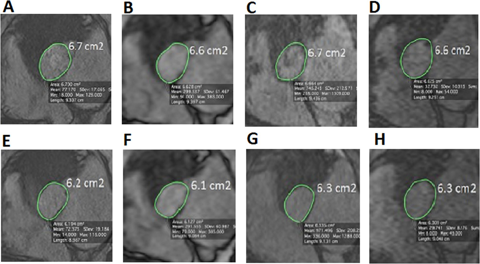
Fig. 4.

Comparison of aortic annulus area between the different MR sequences. Aortic annulus area measured with the different MR sequences in an 80 year old TAVR candidate subject during systole (top images) and diastole (bottom images) using 2D cine b-SSFP (a, b), 3D cine b-SSFP (b, f), navigator triggered 3D b-SSFP MRA (c, g) and k–t accelerated 3D cine b-SSFP (d and h). We can appreciate in this figure the similarity in the aortic annular area value between the k-t accelerated 3D cine b-SSFP and the other conventional MR techniques during both systolic and diastolic phases
