
Fig. 5. CTZ and BIF decrease mitochondrial localization of HK2.
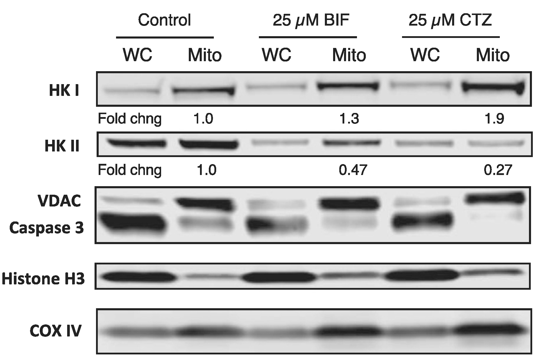
HCT-116 cells were plated and allowed to attach overnight after which cells were left untreated or treated with 25 μM CTZ or BIF for 24 h. Cells were then harvested and whole-cell lysates or mitochondria-enriched fractions were obtained, as outlined in Materials and Methods. Protein was isolated and subjected to PAGE after which the proteins were transferred to a nitrocellulose membrane. The membrane was subsequently probed with antibodies for VDAC1, caspase 3, Cox IV, HK1, HK2 and histone H3. Band intensity was measured on a Li-COR Odyssey scanner. Mitochondrial levels of HK1 and HK2 were normalized to VDAC expression. Fold changes in mitochondrial expression of HK1 and HK2 are compared to levels in untreated cells, which were assigned a value of 1. Results shown are from one of two independent experiments.