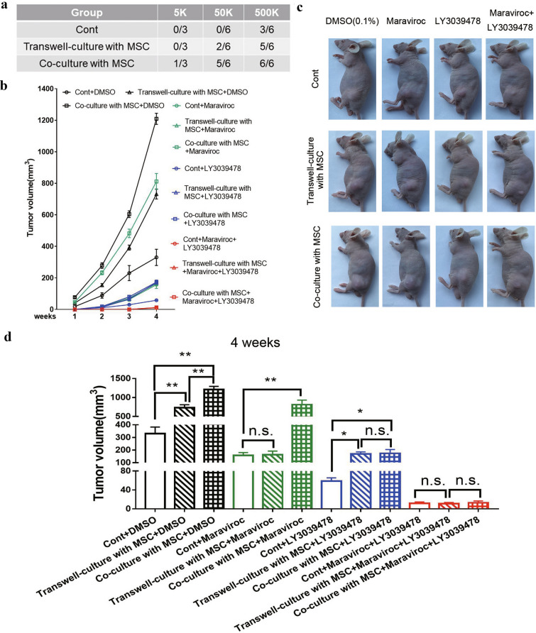
Fig. 5.
MSCs promoted PCa tumor growth by cell–cell contact in vivo via the Notch pathway. a PC-3 cells following mono-culture, transwell-culture, and co-culture with MSCs at indicated cell numbers were subcutaneously transplanted into nude mice (n = 9/group). Tumor formation was monitored at 4 weeks. b Tumor size was measured each week for 4 weeks. Tumor growth curves are shown. c 4 weeks after injection, representative photos showing tumors at the cell injection site were taken in each group. (Scale bar, 100 μ M) d 4 weeks after injection, tumor volume in each group was calculated. Data are presented as the mean ± SD; *P < 0.05, **P < 0.01; ***P < 0.001

