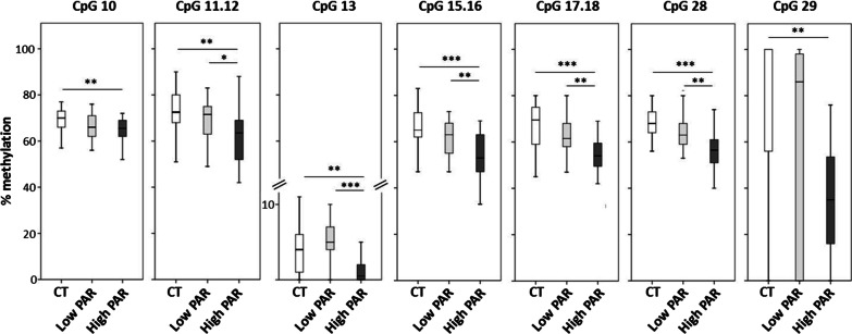
Fig. 5.
Levels of SOCS3 CpG methylation in low and high PAR T2DM patients and in control individuals. Box-whisker plot showing the methylation percentage of different SOCS3 CpG sites as detected by the EpiTYPER assay. Boxes show the median, the 25th and the 75th percentiles. Whiskers show the minimum and the maximum data point. Comparison between groups was performed by the Kruskal–Wallis test followed by the Dunn-Bonferroni post hoc test for pairwise comparisons. Significant differences are indicated by the asterisks. *p ≤ 0.05, **p ≤ 0.01, ***p ≤ 0.001. N = 48 CT, 30 low PAR, 31 high PAR

