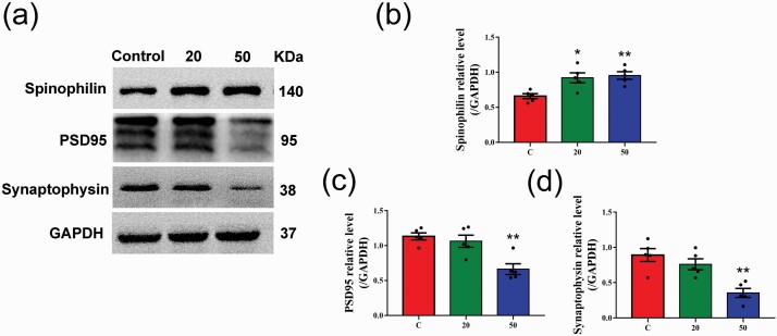
Figure 7.
Effects of gossypol exposure on the expression of synaptic proteins in the hippocampus. (a) Representative western blots of spinophilin, PSD95, synaptophysin, and GAPDH and densitometric quantification of the protein levels in the hippocampus at P21. Relative protein levels of spinophilin (b), PSD95 (c), and synaptophysin (d). Gossypol increased the levels of spinophilin in the hippocampus, while the levels of PSD95 and synaptophysin were decreased. Data represent the mean ± SEM (n = 5 per group, 5 offspring from 5 dams in each group). *P < .05 and **P < .01 compared with the controls.

