Fig. 5.
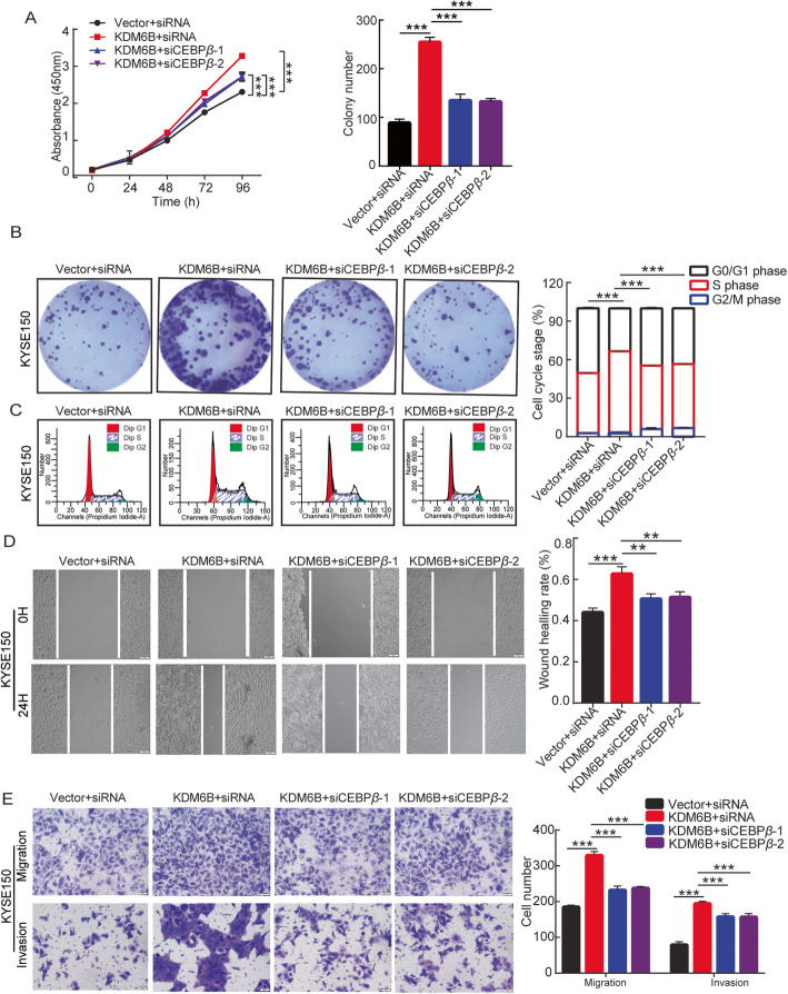
KDM6B-Stimulated proliferation and Metastasis of ESCC Cells through Upregulation of C/EBPβ Expression. KYSE150 cells were successfully cotransfected with KDM6B and C/EBPβ. a The CCK-8 assay was used to determine cell proliferation every 24 h for 4 days. b Representative crystal violet staining of clone formation (left panels). and statistical graphs of Clone numbers (upper right panels). c Representative images of cell cycle distribution (left panels) and statistical graphs of cell cycle changes (right panels). d Cell migration was determined using the wound healing migration assay (magnification, × 100) . Representative images of cells at 0 and 24 h of wound healing assay. Image j was used to analyze the results of wound scratch images. A bar graph shows the quantitative results of scratch healing. e Cell migration or invasion was determined using Transwell migration or invasion assay. A representative images of crystal violet stained for KYSE150 by transwell migration or invasion assays. A statistical graph shows cell number (right panels). Results are representative of three independent experiments and are expressed as the mean ± S.D. * p < 0.05, ** p < 0.01, *** p < 0.001

