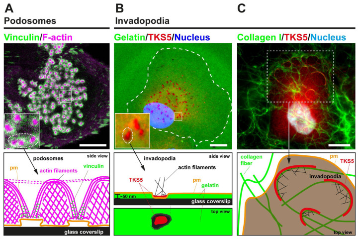
Figure 1. Podosome and invadopodia basics.
(A) The upper image represents a human monocyte-derived immature dendritic cell that was plated for 60 minutes on a glass coverslip, fixed, and stained for actin (magenta) and vinculin (green). A large cluster of dot-shaped podosomes is visible at the adherent surface. The lower panel schematically depicts the side view of a few individual podosomes, highlighting the different actin architectures. (B) In the upper image, MDA-MB-231 cells expressing TKS5GFP were plated for 60 minutes on a thin substratum of fluorescently labeled cross-linked gelatin (pseudocolored in green). Fixed cells were stained for GFP (pseudocolored in red), and the nucleus was stained with DAPI (pseudocolored in blue). Breast cancer cells form typical punctate gelatinolytic invadopodia enriched in the scaffolding protein TKS5. The lower panel schematically depicts a punctate invadopodium with TKS5 enrichment leading to the formation of a small F-actin patch lying on top of a region of gelatin degradation. (C) Upper image: MDA-MB-231 cells expressing TKS5GFP were plated for 60 minutes on a thick layer of fluorescently labeled fibrillar type I collagen (pseudocolored in green). Cells were stained for GFP (pseudocolored in red), and the nucleus was stained with DAPI (pseudocolored in blue). The lower panel schematizes elongated TKS5-, F-actin-rich invadopodia forming at contact sites with the collagen fibers. Scale bars, 10 μm. pm, plasma membrane.

