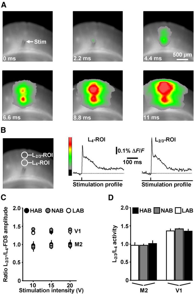
Figure 4.
VSDI analysis of evoked neuronal activity propagations in the motor and visual cortex of HAB, NAB, and LAB mice. A, VSDI filmstrip depicting the spread of neuronal activity triggered by an electrical stimulation pulse (10 V) delivered to L5 of V1 of a LAB animal (for color bar, see B). B, ROI-based extraction of L4 and L2/3 FDSs. C, D, L2/3/L4 activity varied neither in M2 nor in V1 among HAB, NAB, and LAB mice (M2: HAB, n = 8 slices/4 animals; NAB, n = 7 slices/4 animals; LAB, n = 8 slices/4 animals; V1: HAB, n = 7 slices/4 animals; NAB, n = 8 slices/4 animals; LAB, n = 8 slices/4 animals).

