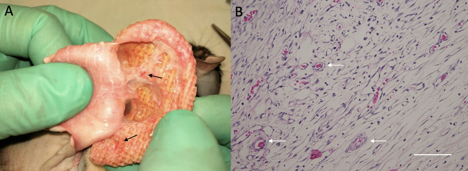
Figure 3:

Images showing evidence of angiogenesis upon harvesting the scaffolds at 8 weeks. A – View demonstrating evidence of angiogenesis on gross examination. B – Representative microscopic image demonstrating vasculature ingrowth on Hematoxylin and Eosin staining, where the scale bar represents 100 microns. Vascularization is noted by the arrows.
