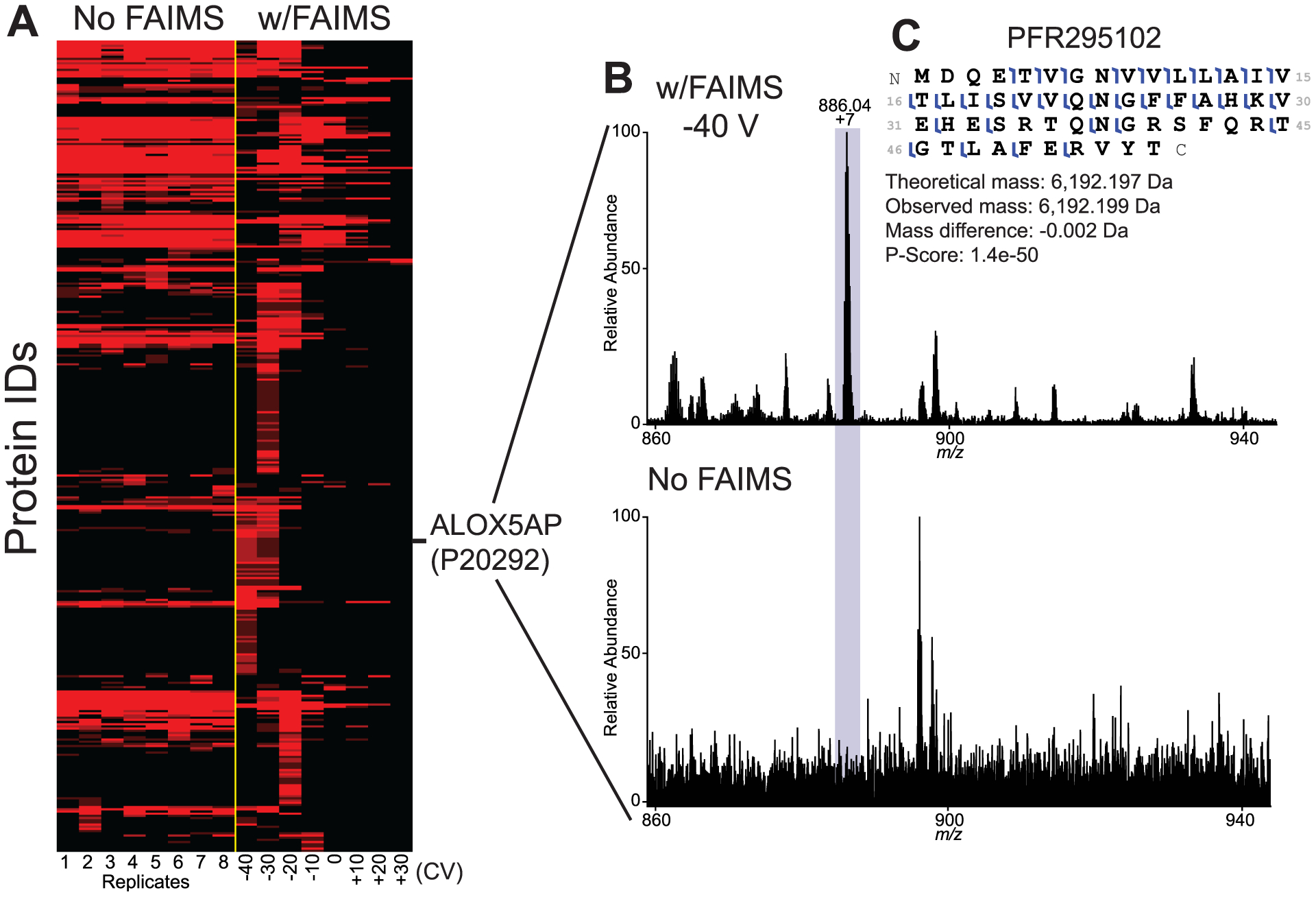
Figure 3.

Changing the compensation voltage (CV) results in detection of a hidden population of proteins through improved signal-to-noise values. (A) Protein-level heatmap of CD3+ T cell proteins identified without FAIMS (8 technical replicates) or with FAIMS applying different CVs from −40 V to +30 V. CVs for injections analyzed with FAIMS are indicated at the bottom of the heatmap. (B) Mass spectrum of proteoform PFR295102 from UniProt accession P20292 (Arachidonate 5-lipoxygenase-activating protein) analyzed with FAIMS (top panel) and without FAIMS (lower panel) at same retention time. (C) Fragmentation map of PFR295102 showing 52% sequence coverage in the sample using FAIMS.
