
Figure 3.
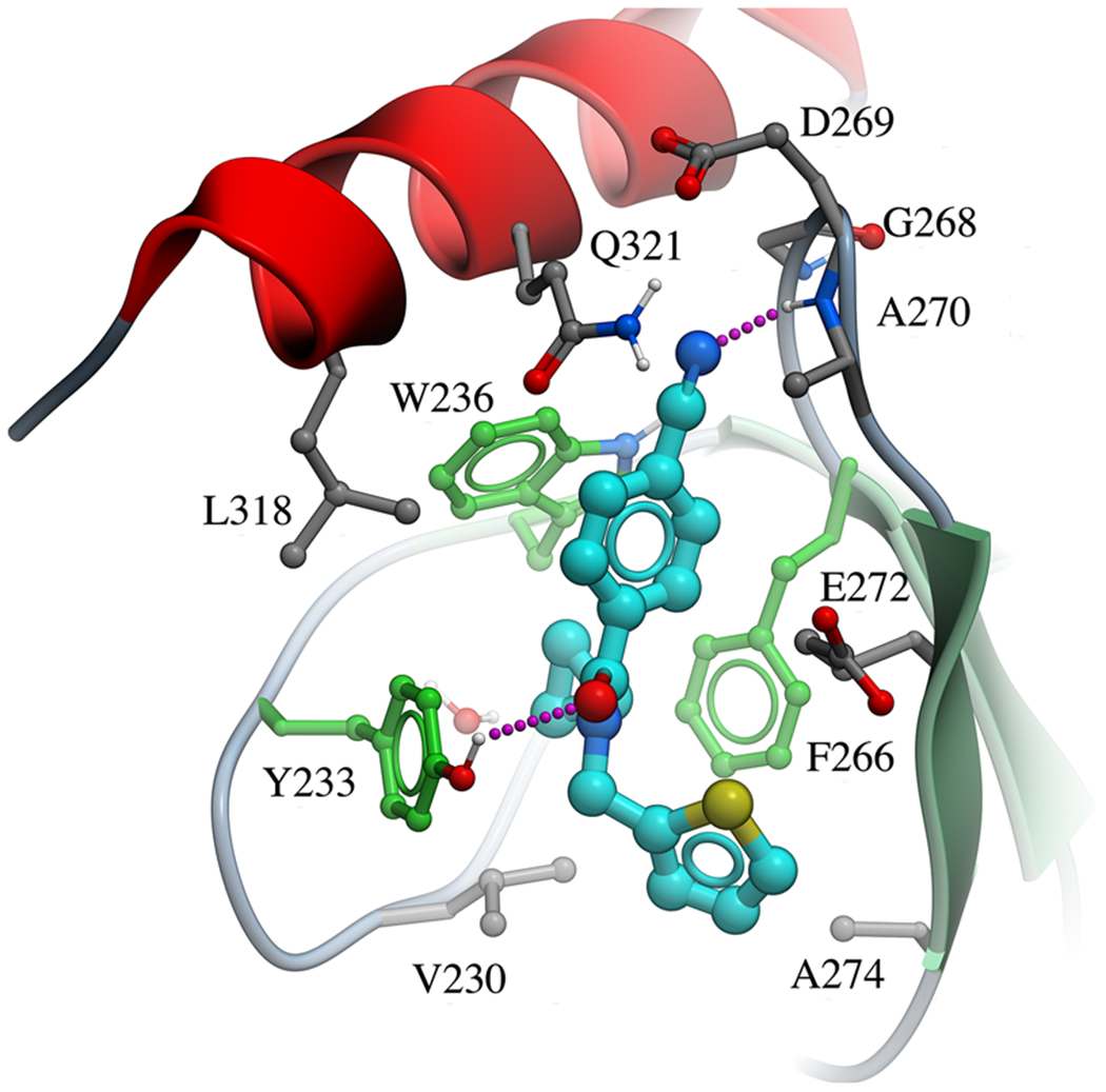
Crystal structure of the NSD2-PWWP1 domain in complex with 3f [PDB code 6UE6]. Binding pocket residues (gray) and the antagonist (cyan) are displayed as sticks and hydrogen bonds as magenta dashed lines. Aromatic cage residues are colored in green.