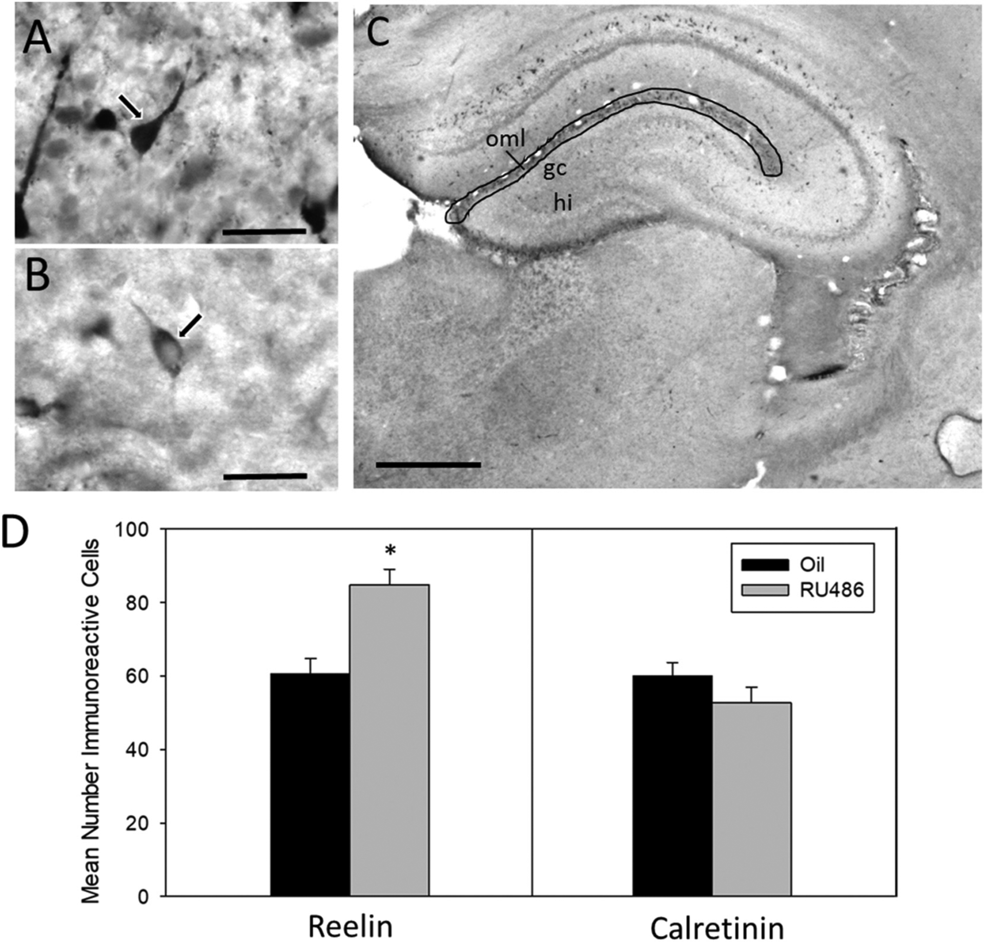
Fig. 4.

Number of reelin and calretinin cells in suprapyramidal outer MOL of P7 males. (A) Representative image of Calretinin-ir cell in suprapyramidal layer of MOL at P7 taken at 100× magnification (scale bar = 30 μm). Arrow illustrates distinctive ‘tadpole-like’ morphology indicative of Cajal-Retzius cells. (B) Representative image of Reelin –ir in suprapyramidal layer of MOL at P7 taken at 100× magnification (scale bar = 30 μm). (C) Representative image of dorsal hippocampus indicating region of quantification of reelin and calretinin-ir (gc (granule cell layer), hi (hilus), oml (outer molecular layer)) (scale bar = 500 μm). (D) Mean number (±sem) of reelin immunoreactive (ir) cells and calretinin-ir cells in the suprapyramidal outer molecular layer of the dentate gyrus at postnatal day 7 in RU486- or oil-treated male rats (*significantly different from controls, p < 0.05).
