1. Introduction
Recently, steady-state and time-resolved fluorescence properties of Hoechst 33342 [bis-benzimide, 2,5′-bi-l H-benzimidazole, 2′-(4-ethoxyphenyl)-5-(4-methyl-piperazinyl)] were examined for one-photon (OPE) and two-photon (TPE) excitation [1]. The Hoechst 33342 (HOE) stain binds to DNA with a significant increase in fluorescence. Consequently, HOE is widely used for selective visualization of DNA in fluorescence microscopy, flow cytometry and chromosome analysis. HOE was found to display a large cross section for TPE within the fundamental wavelength range of pyridine 2 and rhodamine 6G dye lasers, i.e. 690 to 770 nm and 560 to 630 nm, respectively.
As recently demonstrated, the steady-state limiting anisotropy by photoselection of luminescent molecules (LM) in isotropic rigid media is markedly higher in two-photon than in one-photon excitation [2–4]. As compared to that latter, two-photon excitation of LM embedded in anisotropic media (stretched PV film) results in the enhanced photoselection [5].
The aim of this work is to investigate the fluorescence emission anisotropy (r) of HOE in anisotropic (stretched) poly(vinyl alcohol) (PVA) films for OPE and TPE, and to determine the absolute direction of the absorption transition moment (with respect to the long molecular axis) of a prolate HOE molecule and the angle between the directions of absorption and fluorescence transition moments.
2. Theoretical Background
As shown in [5], the fluorescence emission anisotropy in the case of linearly polarized exciting light is given by (see Fig. 1 in [5]):
| (1) |
where
| (2) |
applies to the general case of multi-photon (n) excitation. Rs is the stretch ratio and β is the angle between the directions of absorption and emission transition moments. β is constant for a given transition in the LM.
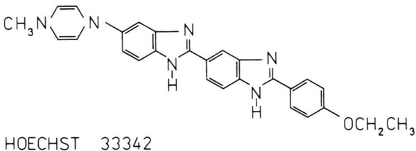
Fig. 1.
Structural formula of Hoechst 33342 (HOE).
In (2), in view of the polarized absorption, the direction distribution in the excited state for rod-shaped LM was used in the form
where
is the Tanizaki [6] distribution function for molecules in the ground state.
By substituting in (2) one obtains
| (3) |
where
| (4) |
For (isotropic rigid solution) a = 0, (3) implies
| (5) |
| (6) |
Thus, for one-photon absorption (n = 1), the wellknown Perrin equation [7, 8] is obtained, while for two-photon absorption (n = 2) one obtains the equation found in [2–4].
In general, according to (3), for Rs > 1 and one-photon absorption (n = 1) one obtains
| (7) |
and for two-photon absorption (n = 2)*
| (8) |
The orientation degree of the environment (e.g. the film stretch ratio Rs) was assumed to be a measure of the arrangement of the emitted molecules. In practice, the molecular arrangement is essentially affected by the shape of the molecule [9] (a part originating from the stretch ratio, Rs). As shown in [9], the change in the emission anisotropy due to stretching of the polymer film can be investigated as a function of the measured dichroic ratio (A∥ and A⊥ are the components of the absorbance, A = ε c l, parallel and perpendicular to the stretching direction (the z-axis) of the polymer film, respectively, ε is the molar absorption coefficient in litres per mol · cm, c is the concentration in mol/litre, and l is the length in cm), which is the actual degree of the orientation of long molecular axes, taking also into account such factors as the degree of the orientation of the polymer, the shape factors, etc.
For linear symmetrical molecules, when the absorption transition moment lies along the long molecular axis (φ = 0), the dependence of the dichroic ratio, Rd, on the stretch ratio, Rs, is linear and can be described by [9]
| (9) |
Hence, one obtains
| (10) |
and (4) becomes
| (11) |
3. Experimental
Hoechst 33342 (Fig. 1) from Molecular Probes was used without further purification. Isotropic films were made of a 15% aqueous solution of poly(vinyl alcohol) (PVA) in which the HOE molecules were set by methanol. PVA films were stretched at about 350 K, with controlled stretching rate. The preparation method was described in detail in [10, 11]
The polarized absorption, fluorescence and emission anisotropy measurements were carried out by the apparatus described in [1, 4, 12].
4. Results and Discussion
4.1. Emission Spectra of HOE in PVA Film
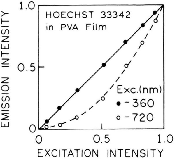
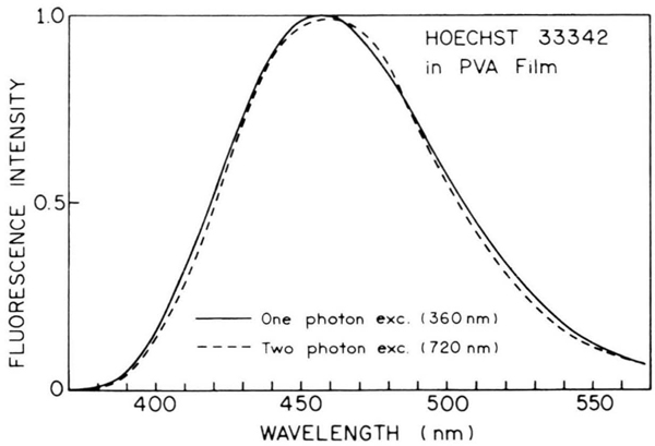
Figure 2 shows the dependence of the observed fluorescence intensity on the excitation intensity for OPE and TPE of HOE in an isotropic PVA film. The linear and quadratic dependences observed for the 360 and 720 nm excitation, respectively, demonstrate that the long wavelength-induced emission signal is due to a biphotonic process. The emission spectra of HOE in PVA film at room temperature are shown in Figure 3. The same emission spectra were observed for OPE and TPE at the 360 and 720 nm excitation, respectively. The fact that the emission spectra are identical for OPE and TPE indicates that emission occurs from the same state, irrespective of the mode of excitation. Similar results were obtained for HOE in ethanol [1].
Fig. 2.
The dependence of the emission intensity of HOE in PVA film at 20 °C on the excitation intensity for one-photon (●) and two-photon (○) excitation.
Fig. 3.
Normalized fluorescence of HOE in PVA film at 20°C with one-photon and two-photon excitation.
4.2. Dichroism of HOE in Stretched PVA Films
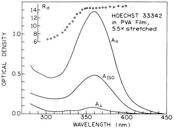
The absorption spectra of HOE, measured in PVA film 5.5-fold stretched for the absorbance components, A∥ and A⊥, and the dichroic ratio, versus wavelength are shown in Figure 4.
Fig. 4.
Optical density components (absorbances), A∥, A⊥ and AISO, and the ratio of HOE in a 5.5-fold stretched PVA film.
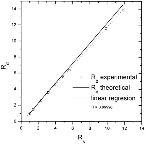
The elongated HOE molecule was found to become very well oriented during the stretching of the PVA film. Above 340 nm, the dichroic ratio, Rd, in the longwave absorption band remains constant. Figure 5 shows the dependence of Rd on the stretch ratio, Rs, of PVA film. For a perfectly linear molecule, according to (9), a linear dependence can be expected. Above Rs = 6, the measured experimental points deviate only slightly from the straight line (solid line) given by (9). Hence, the absorption transition moment lies along the long axis of the HOE molecule (i.e. angle φ is zero; Fig. 1 in [9]).
Fig. 5.
Theoretical dependence of the dichroic ratio Rd on the stretch ratio Rs for φ = 0° ((9) and (4) in [9]); - experimental points.
4.3. Fluorescence Anisotropy of HOE in Stretched PVA Films
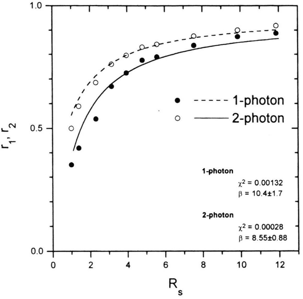
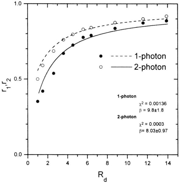
When the angle β between the directions of absorption and emission transition moments is zero, the theoretical fundamental values of emission anisotropy rf for OPE and TPE are 2/5 and 4/7, respectively, (6). The measured limiting fluorescence anisotropies, r0, for HOE in isotropic PVA film are smaller and amount to 0.351 and 0.5 for OPE and TPE, respectively. The values of r measured for HOE in stretched PVA films, shown in Figs. 6 and 7, are compared with theoretical curves, (1), (7) and (8), for OPE and TPE versus Rs = k3/2 (k is the multiplication factor of stretching [10]) and Rd, respectively. Based on the best fit, the angle β between the directions of absorption and fluorescence transition moments can be determined.
Fig. 6.
Dependence of r1 and r2 on the degree of stretching, Rs, for HOE in PVA films for OPE (●) and TPE (○) excitation.
Fig. 7.
Dependence of r1 and r2 on the dichroic ratio for HOE in PVA films for OPE (●) and TPE (○) excitation.
The replacement of Rs with Rd according to (10), in the case considered has no great significance since the determined values of angles β are only slightly different. For TPE, the angle β is smaller, amounting to about 8°. A better agreement is observed between the measured values and the theoretical curve calculated from (8). For low Rs and Rd values (to about 3), i.e. weakly stretched PVA film, the observed emission anisotropy is underrated, which can be accounted for by the fact that in larger polymer cavities there is a better libration of HOE molecules. As the film is stretched, the cavities around the HOE molecules elongate, thus hindering the molecular motions.
A separate problem is the lower value of the limiting emission anisotropy, r0, compared to the fundamental emission anisotropy, rf, of the isotropic solution, 2/5 and 7/4 for OPE and TPE, respectively. The small difference between the fundamental and the limiting anisotropies, rf — r0, measured with the steady-state technique, can be due to vibrations performed by an LM following excitation as a result of the initial shock [8, 13]. For complex LM, the deformation vibrations spoil the molecular configuration and the link between the transition dipole moments and the molecule loses its rigidity.
Acknowledgments
It was found by investigating dichroism and emission anisotropy in the case of one- and two-photon excitation of Hoechst 33342 [bis-benzimide,2,5′-bi-lH-benzimidazole, 2′-(4-ethoxyphenyl)-5-5(4-methyl-l-piperazinyl)] in stretched poly(vinyl alcohol) (PVA) films, that the absorption and fluorescence transition moments lie along the long molecular axis of the molecule studied. The slight deviation of the transition moment direction in fluorescence (about 8°) from that in absorption can be due to the incomplete linearity of the Hoechst molecule.
Dedicated to Professor Gregorio Weber in the occasion of his 80th birthday
Footnotes
References
- [1].Gryczyński I and Lakowicz JR, J. Fluoresc. 4, 331 (1994). [DOI] [PubMed] [Google Scholar]
- [2].Lakowicz JR, Gryczyński I, Gryczyński Z, Danielsen E, and Wirth MJ, J. Phys. Chem. 96, 3000 (1992). [DOI] [PMC free article] [PubMed] [Google Scholar]
- [3].Lakowicz JR, Gryczyński I, and Danielsen E, Chem. Phys. Lett. 191, 47 (1992). [DOI] [PMC free article] [PubMed] [Google Scholar]
- [4].Kawski A, Gryczyński I, and Gryczyński Z, Z. Naturforsch. 48 a, 551 (1993). [Google Scholar]
- [5].Kawski A and Piszczek G, Z. Naturforsch. 49 a, 936 (1994). [Google Scholar]
- [6].Tanizaki Y, Bull. Chem. Soc. Japan 38, 1798 (1965). [Google Scholar]
- [7].Perrin F, Ann. Physique 12, 169 (1929). [Google Scholar]
- [8].Kawski A, Critical Rev. Anal. Chem. 23, 459 (1993). [Google Scholar]
- [9].Gryczyński Z and Kawski A, Z. Naturforsch. 42 a, 1396 (1987). [Google Scholar]
- [10].Kawski A and Gryczyński Z, Z. Naturforsch. 41a, 1195(1986). [Google Scholar]
- [11].Kawski A, Developments in Polarized Fluorescence Spectroscopy of Ordered Systems, in: Optical Spectroscopy in Chemistry and Biology – Progress and Trends (Fassler D, ed.), VEB Deutscher Verlag der Wissenschaften, Berlin: 1989. [Google Scholar]
- [12].Kawski A, Gryczyński Z, Gryczyński I, and Kuśba J, Z. Naturforsch. 47 a, 471 (1992). [Google Scholar]
- [13].Jabloński A, Acta Phys. Polon. 26, 427 (1964); 28, 717 (1965). [Google Scholar]


