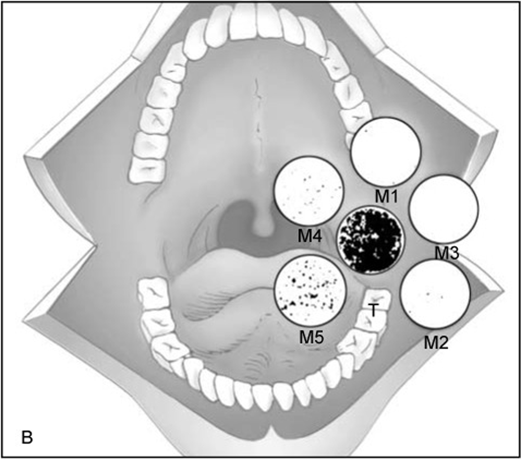
Figure 1.
Figure adapted from Brennan et al. T represents the primary tumor and M1-M5 represent margins. Mutant specific oligomers were identified for each patient and oligonucleotide probes were hybridized with phosphorous-32 combined with DNA extracted from margin tissue. Radiographs were obtained with hybridizing plaques being radio-opaque and representing presence of p53 mutation. 38% of patients with positive p53 margins recurred locally compared to 0% of those with negative p53 margins. RightsLink license number 4870271313736.

