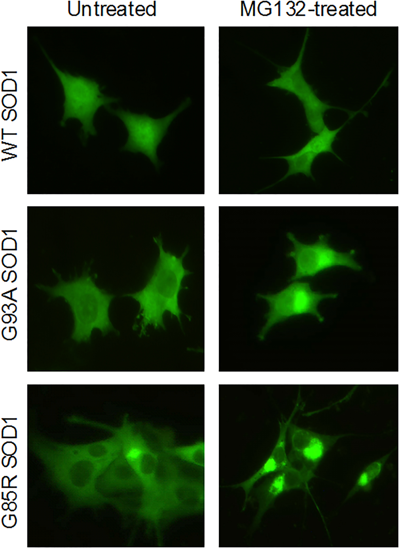
Figure 1. Mutant but not wild type SOD1 forms protein aggregates in cells treated with the proteasome inhibitor MG132.

Fluorescence micrographs of PC12 cells expressing YFP tagged wild type (WT), G93A mutant (G93A) and G85R mutant (G85R) SOD1 proteins. The micrographs show the effect of treating cells with 200 nM MG132 for 24 h. The wild type SOD1 cells are unaffected while cells expressing mutant SOD1 show an elevated fraction of cells with large peri-nuclear aggregates.
