
Figure 1.
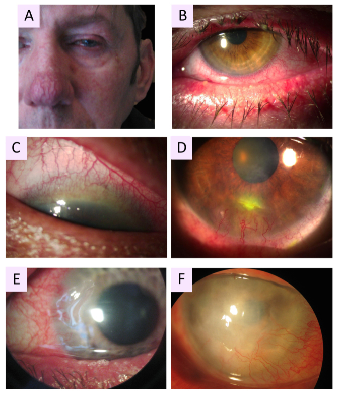
Clinical manifestations of oculocutaneous rosacea. A: Patient with phymatous rosacea–associated rhinophyma, blepharophyma, nasal and facial erythema with telangiectasia. B: Blepharophyma with thickened lid edges, lid margin telangiectasia, meibomian gland dysfunction (MGD). C: Corneal neovascularization (CNV) of ocular rosacea growing from the superior limbus with a crescent pattern forming a vascular pannus. D: Catarrhal corneal infiltrate caused by rosacea. E: Typical peripheral ulcerative keratitis (PUK) of rosacea, corresponding to sterile corneal melting of a crescentic area with newly formed stromal vessels. F: Advanced stage of ocular rosacea with white corneal infiltrates and whole corneal neovascularization, including the visual axis.