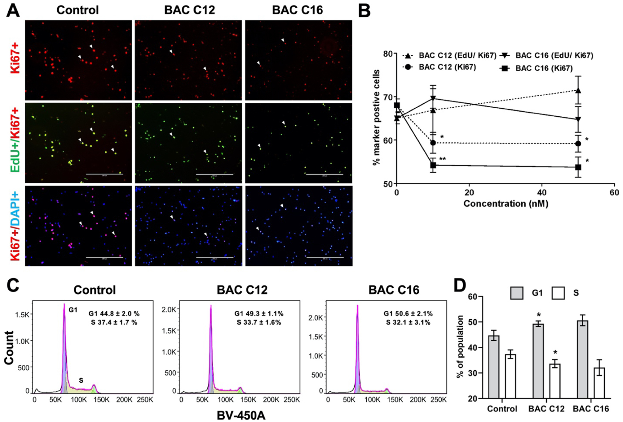
Figure 3. BACs decrease pool of proliferative NPCs.

(A) Immunocytochemistry for Ki67 (red) and EdU (green), counterstained with DAPI (blue) of NPCs from dissociated neurospheres exposed to vehicle control (0 nM) or 50 nM of BAC C12 or BAC C16 from DIV 4 to DIV 7. (B) Quantification of proliferation rate (Edu/Ki67) and percentage of proliferative cells (Ki67) cells. Adjusted P value: *, P < 0.05; **, P < 0.01; ***, P < 0.001. (C) Cell cycle analysis of nuclei isolated from dissociated neurospheres exposed to vehicle control or 50 nM of BAC C12 or BAC C16 from DIV 4 to DIV 5. (D) Quantitation of population of cells in G1 or S phase in (C); *, P < 0.05 relative to Control. N = 4 biological replicates per condition.
