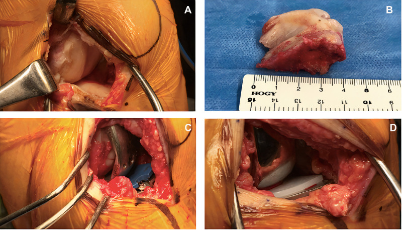
Figure 3.
(A) Perioperative photographs documenting the extent of cartilage damage at the lateral aspect of the right knee. (B) Excised portion of the proximal tibia with dimensions approximately 40 × 30 × 20 mm. (C) Placement of the trial implant with the smallest available polyethylene spacer insert. (D) Final UKA implant with cement and polyethylene insert as shown.

