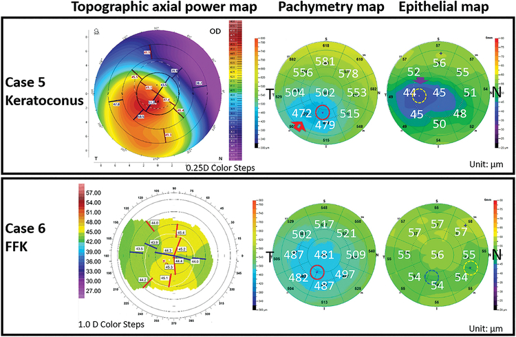
Figure 6.
Examples of misclassified keratoconic cases. Both eyes were suspicious for keratoconus according to decision tree step 1 output. But their thickness map patterns did not meet the criteria for keratoconus in step 2 of the decision tree. A keratoconic eye (Case 5, top row) was misclassified by the decision tree because the thinnest cornea and epithelium were separated by more than one map zone. Both pachymetric and epithelial thickness maps demonstrated a clear concentric thinning pattern (arrows). An FFK eye (Case 6, bottom row) was misclassified by the decision tree due to 2 sectors separating the thinnest cornea (red circle) and epithelium (yellow dotted circle). There was a second area of thin epithelium (blue dotted circle) that was co-located with the thinnest cornea.

