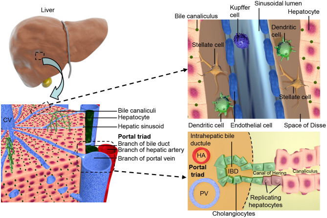
Figure 1.
Shows the structural organization of the liver at different scales. The two lobes of the liver consist of hexagonal units known as lobules, these consist of a central vein surrounded by portal triads resulting in an axis, referred to as liver zonation, along which hepatocyte and sinusoidal function varies. The portal triads consist of mesenchymal cells surrounding the portal vein (PV), hepatic artery (HA), and the cholangiocyte lined intra-hepatic bile ducts (IBD). These are joined to the hepatic canaliculi by the Canals of Hering. The microvasculature of the liver, known as the sinusoid, interfaces the blood supply with the hepatocytes and is also the location of the hepatic stellate cells and Kupffer cells.

