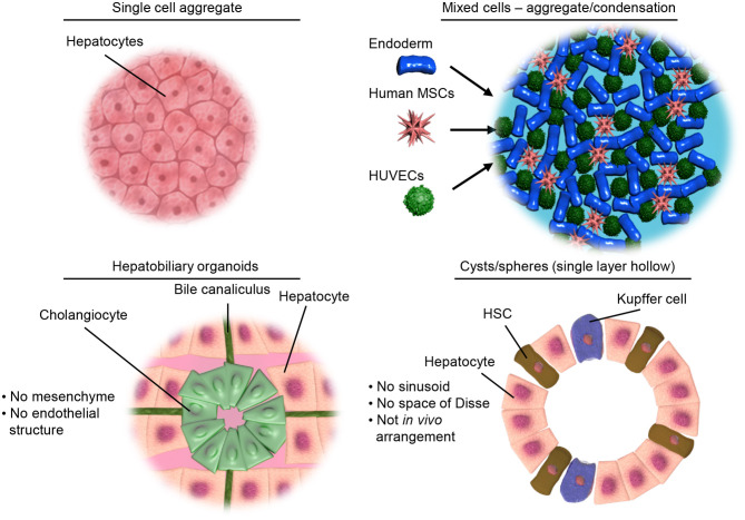
Figure 2.
Illustrative examples of different types of 3D culture models. Top left, showing a simple aggregate of a single cell type. Top right, showing aggregates of mixed cell types but limited structural organization as achieved by condensation of pre-differentiated cells. Bottom left, representing the hepatobiliary organoids recapitulating the structure and interactions of the liver parenchymal cells. Bottom right, showing organoids containing both parenchymal and non-parenchymal cells of the liver necessary for modeling inflammation and liver disease.

