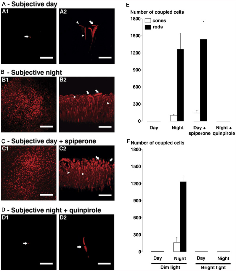
Figure 11.
The circadian clock in the goldfish retina controls rod-cone tracer coupling by activating dopamine D4 receptors in the day but not at night. (A–D) Following iontophoresis of biocytin into individual cones in intact neural goldfish retina, tracer remained in a few cells (indicated by arrows) near the injected cone during the subjective day (A) and during the subjective night in presence of D4 receptor agonist quinpirole (1 μM, D). It diffused into many rods and cones during subjective night (B) and during subjective day in the presence of D4 receptor antagonist spiperone (10 μM, C). In each of (A–D), confocal images of a whole-mount retina at the level of the rod inner segments are shown on the left (A1–D1) and perpendicular views of the 3-D reconstruction of the photoreceptor cells from the same retina are shown on the right (A2–D2). Some cones and rods are indicated by arrows and arrowheads, respectively. Scale bars (A–D): 50 μm. (E,F) Average numbers of stained cones (open bars) and rods (filled bars) following biocytin injections into individual cones (1 cone injected/retina) under dark-adapted (E) and light-adapted (F) conditions are shown. (E) Under dark-adapted conditions (>60 min), the number of tracer coupled rods and cones was significantly greater during the night and during the day following spiperone treatment than during the day under control conditions. (F) Under dim light-adapted conditions (i.e., −5 logIo, 500 ms light flashes at 0.125 Hz for >60 min), the number of tracer-coupled rods and cones was significantly greater during the night compared to the day (i.e., results are similar to those obtained in the dark). In contrast, under bright light-adapted conditions (i.e., −2 logIo, 500 ms light flashes at 0.125 Hz for >60 min) in day and night, biocytin was restricted to the injected cone; no other cells were labeled. Error bars represent SEM. Modified from Ribelayga et al. (2008).

