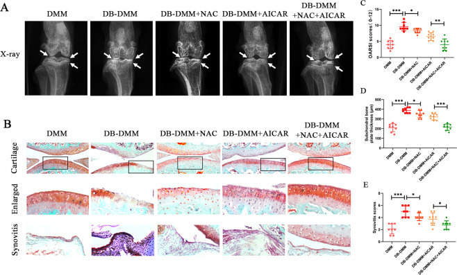
Fig. 6. NAC and AICAR ameliorate DB-accelerated OA development in a DB-DMM mouse model in vivo.
A Digital X-ray image of mice knee joints in the various experimental groups. Joint space, calcification of the cartilage surface, and osteophyte formation are represented by white arrows. B Representative Safranin-O staining of cartilage and synovitis in various experimental groups at 4 weeks post surgery. C OARSI scores of cartilage. D. Subchondral bone plate thicknesses. E Synovitis scores. Data are presented as means ± SD (n = 8). *P < 0.05, **P < 0.01 and, ***P < 0.001.

