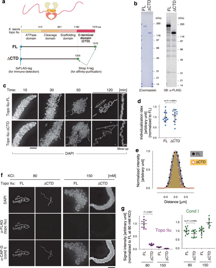
Fig. 2. Topo IIα-ΔCTD is proficient in chromatid individualization but is deficient in chromatid thickening.
a Schematic presentation of structures of full-length (FL) and CTD-deleted (ΔCTD) versions of recombinant Xenopus laevis topo IIα. b Purified topo IIα-FL and topo IIα-ΔCTD were analyzed by SDS-PAGE and stained with Coomassie Blue. The same set of samples was also subjected to immunoblotting using anti-FLAG antibodies. This experiment was repeated three times with similar results. c–e Xenopus sperm nuclei were incubated in the reconstitution reaction mixture containing either topo IIα-FL or topo IIα-ΔCTD. At the indicated time points, the resultant chromatin was fixed and stained with DAPI. Blow-up images of cropped parts (indicated by the dashed rectangles in the original 120-min images) are shown on the right (c). Individualization indices at 120 min are plotted. The mean ± s.d. is shown (n = 12 clusters of chromatids). P values were assessed by two-tailed Welch’s t-test (ns not significant) (d). Profiles of normalized signal intensities of DAPI along lines drawn perpendicular to chromatid axes were analyzed. The mean ± s.d. is shown (n = 15 lines from 5 chromatids) (e). f, g Chromatid reconstitution assays were performed with topo IIα-FL or topo IIα-ΔCTD in a buffer containing 80 mM or 150 mM KCl. After a 150-min incubation, the resultant structures were fixed and processed for immunolabeling (f). Signal intensities of topo IIα and CAP-G on chromatids were analyzed. The mean ± s.d. is shown (n = 9 clusters of chromatin). P values were assessed by two-tailed Welch’s t-test (ns not significant) (g). Bars, 5 µm.

