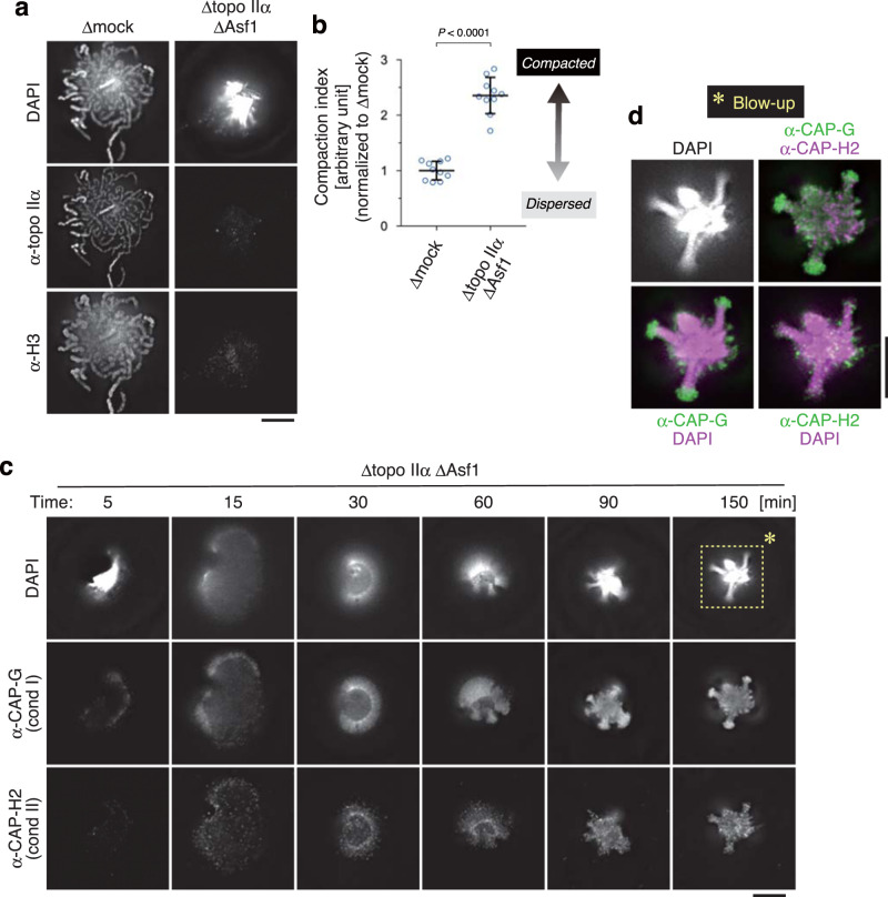
Fig. 4. An unusual chromatin structure is produced in the cell-free extract depleted of both topo IIα and Asf1.
a, b Mouse sperm nuclei were incubated in a control extract (Δmock) or an extract depleted of both topo IIα and Asf1 (Δtopo IIα ΔAsf1) for 150 min and labeled with antibodies against topo IIα and histone H3. DNA was counterstained with DAPI (a). The compaction indices (the average DAPI intensities per unit area) are plotted. The mean ± s.d. is shown (n = 10 clusters of chromatin). P values were assessed by two-tailed Welch’s t-test (b). c, d Mouse sperm nuclei were incubated in the Δtopo IIα ΔAsf1 extract at 22 °C. At the indicated time points, the reaction mixtures were fixed and labeled with antibodies against CAP-G (a condensin I subunit) and CAP-H2 (a condensin II subunit). DNA was counterstained with DAPI. This experiment was repeated three times with similar results (c). Blow-up images of the cropped part (indicated by the dashed rectangle in the original DAPI image at 150 min) are shown in grayscale (DAPI) and pseudo-colors (merged images for the indicated combinations) (d). Bars, 5 µm.

