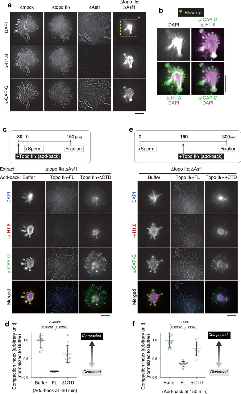
Fig. 5. Topo IIα-FL, but not topo IIα-ΔCTD, can efficiently rescue the defects observed in the extracts depleted of both topo IIα and Asf1.
a, b Mouse sperm nuclei were incubated in a control extract (Δmock) or an extract depleted of either topo IIα (Δtopo IIα), Asf1 (ΔAsf1), or both (Δtopo IIα ΔAsf1). After a 150-min incubation at 22 °C, the resultant structures were labeled with antibodies against the linker histone H1.8 and CAP-G. DNA was counterstained with DAPI. This experiment was repeated three times with similar results (a). Blow-up images of the cropped part (indicated by the dashed rectangle in the original DAPI image in a Δtopo IIα ΔAsf1 extract) are shown in grayscale (DAPI) and pseudo-colors (merged images for the indicated combinations) (b) c, d An extract depleted of topo IIα and Asf1 (Δtopo IIα ΔAsf1) was supplemented with either buffer, topo IIα-FL, or topo IIα-ΔCTD. After a 30-min incubation at 22 °C, mouse sperm nuclei were added to these extracts and incubated for another 150 min. The resultant chromatin structures were fixed and processed for immunolabeling with the antibodies indicated (c). The compaction indices were analyzed and are shown in Fig. 4b. The mean ± s.d. is shown (n = 12 clusters of chromatin). P values were assessed by two-tailed Welch’s t-test (d). e, f Mouse sperm nuclei were first incubated in a Δtopo IIα ΔAsf1 extract to allow sparkler formation. At 150 min, the reaction mixtures were supplemented with either buffer, topo IIα-FL, or topo IIα-ΔCTD. After another 150-min incubation at 22 °C, the samples were fixed and processed for immunolabeling (e). The compaction indices were analyzed and are shown as above. The mean ± s.d. is shown (n = 12 clusters of chromatin). P values were assessed by two-tailed Welch’s t-test (f). Bars, 5 µm.

