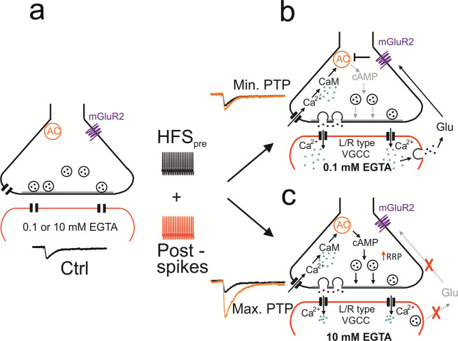
Fig. 5. Mechanisms of PTP and possible signaling pathways underlying transsynaptic modulation at the MFB–CA3 pyramidal neuron synapse.
a MFB–CA3 synapse during resting conditions. Presynaptic terminal in black on top, postsynaptic compartment in red at the bottom. AC stands for adenylyl cyclase, mGluR2 indicates extrasynaptic metabotropic glutamate receptor 2 (and mGluR3 expressed at lower levels). b MFB–CA3 synapse after tetanic stimulation, leading to postsynaptic activity, in the presence of low (0.1 mM) concentrations of the Ca2+ chelator EGTA in the postsynaptic cell. CaM stands for calmodulin, VGCC for voltage-gated Ca2+ channels, and Glu for glutamate. Ca2+ ions indicated as green dots, glutamate molecules as black dots. Note that in this scenario tetanic stimulation does not trigger PTP, due to retrograde vesicular glutamate signaling. c Same as for b but in the presence of high concentration (10 mM) of the Ca2+ chelator EGTA in the postsynaptic cell. Note that in this scenario tetanic stimulation does trigger PTP, due to the lack of retrograde vesicular glutamate signaling. Transsynaptic modulation of PTP induction may not only occur with the exogenous Ca2+ chelator EGTA, but also with endogenous Ca2+ buffers expressed in the postsynaptic neurons. This may convey both anti-associativity and target cell-specificity to the PTP induction mechanism51.

