Figure 1.
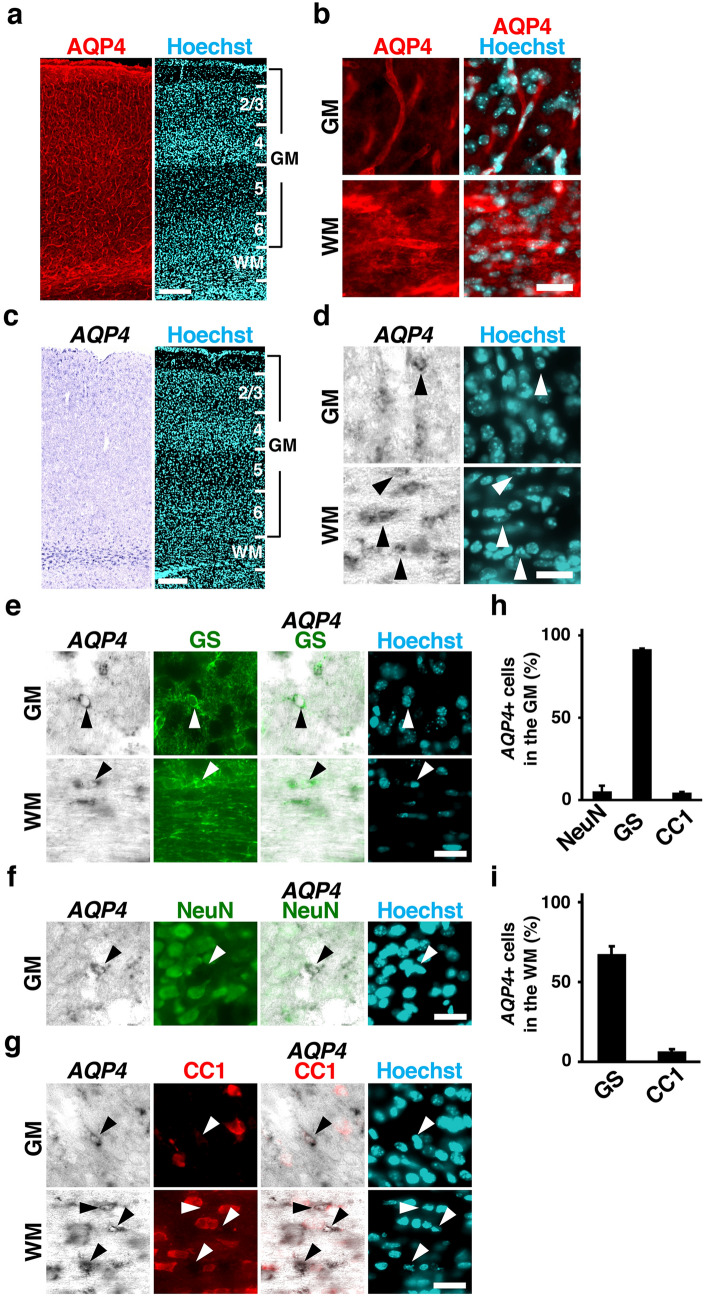
Expression patterns of AQP4 in the developing mouse cerebral cortex. (a,b) Sections of the mouse cerebral cortex at P16 were subjected to AQP4 immunohistochemistry and Hoechst 33342 staining. Low magnification images (a) and high magnification images (b) corresponding to the gray matter (GM) and the white matter (WM) are shown. AQP4 was expressed throughout the developing mouse cerebral cortex. The expression level of AQP4 was higher in the WM than in the GM. (c,d) Sections of the mouse cerebral cortex at P16 were subjected to in situ hybridization for AQP4 and Hoechst 33342 staining. Low magnification images (c) and high magnification images (d) corresponding to the GM and the WM are shown. AQP4 mRNA was preferentially distributed in the WM but was also observed in the GM. (e–g) Sections of the mouse cerebral cortex at P16 were subjected to in situ hybridization for AQP4 and immunohistochemistry for either GS (e), NeuN (f) or CC1 (g). High-magnification images of the GM and the WM are shown. AQP4 was preferentially expressed in GS-positive cells (e, arrowheads), while AQP4 signals were not colocalized with NeuN-positive cells or CC1-positive cells (f and g, arrowheads). (h,i) The percentages of AQP4-positive cells co-expressing NeuN, GS, and CC1 in the GM (h) and the WM (i). n = 3 animals for each condition. Bars represent mean ± SD. Numbers indicate the corresponding layers in the cerebral cortex. GM, gray matter; WM, white matter. Scale bars 200 µm (a,c) and 25 µm (b,d–g).

