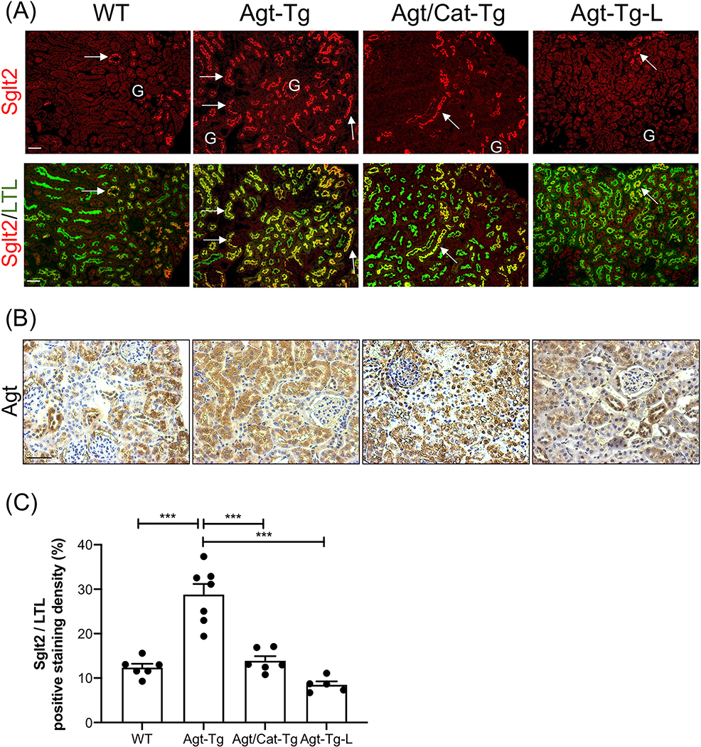
Figure 3: Overexpression of Agt stimulates Sglt2 expression in proximal tubules.
(A) Double immunostaining of Sglt2 (red color, arrow) and a proximal tubular marker (lotus tetragonolobuslectin, LTL) (green color) in WT, angiotensinogen-transgenic (Agt-Tg), angiotensinogen/catalase-transgenic (Agt/Cat-Tg), and Agt-Tg mice treated with losartan (Agt-Tg-L) kidneys (x100). G, glomerulus. (B) Representative immunostaining for Agt in WT, Agt-Tg, Agt/Cat-Tg, and Agt-Tg treated with losartan (×200). (C)Semi-quantification of Sglt2/LTL immunostaining ratio. Scale bars = 50 μm.

