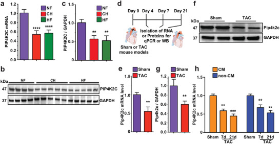
Figure 1.

PIP4K2C and Pip4k2c expression decreases in failing hearts of humans and mice, respectively. a) PIP4K2C mRNA expression in human non‐injured normal left ventricle (LV) (non‐failing, NF, n = 5), cardiac hypertrophy (CH, n = 5), and heart failure (HF, n = 5). b. Western blot of PIP4K2C expression in human LV samples with GAPDH as control (n = 4 for NF, n = 5 for CH, n = 5 for HF). c) Quantitative analysis of b. d) Experimental timeline to analyze Pip4k2c expression in sham‐operated or TAC mouse model. e) mRNA expression of Pip4k2c in WT mouse heart samples compared to those from TAC recipients, 4 days after injury (n = 4). f) Western blot evaluation of Pip4k2c protein in WT mouse heart samples compared to those from TAC recipients 4 days after injury, with GAPDH as a control housekeeping gene. g) Quantitative analysis of Pip4k2c western blot from f (n = 2). h) Pip4k2c mRNA expression in CM or non‐CMs after Sham or 7 or 21 days post TAC injury (n = 3). One‐way ANOVA, Tukey's Multiple Comparison Test were used in a,c; unpaired two‐tailed t‐test was used in e,g; and two‐way ANOVA and Bonferroni post‐hoc tests were used in h. ****, P < 0.0001, ***, P < 0.001, **, P < 0.01.
