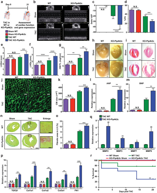
Figure 2.

Loss of Pip4k2c enhances formation of cardiac hypertrophy and fibrosis post TAC injury. a) Experimental timeline to evaluate cardiac function and outcome in Pip4k2c−/− (KO‐Pip4k2c) and Pip4k2c+/+ littermate controls (WT) in a TAC mouse model. b) Representative echocardiography image of left ventricle 21 days post sham or TAC injury in WT or KO‐Pip4k2c. c–f) Echo evaluation of delta % left ventricular ejection fraction (c), fractioning shorting (d), LVIDd (e), and LVIDd (f) 21 days post sham or TAC injury in WT or KO‐Pip4k2c (n = 10). g) Heart weight to tibia length 21 days post sham or TAC injury in WT or KO‐Pip4k2c (n = 10). h,i) Representative images of whole heart (h) and H&E staining (i) 21 days post sham or TAC injury in WT or KO‐Pip4k2c. j) Representative images of wheat germ agglutinin (WGA) staining to evaluate CM size (cross‐sectional area) 21 days post sham or TAC injury in WT or KO‐Pip4k2c. k) Quantitative analysis of j (n = 8). l,m) qPCR analysis of hypertrophic markers 21 days post sham or TAC injury in WT or KO‐Pip4k2c (n = 5). n) Representative images of Sirius red / fast green indicating fibrotic area 21 days post sham or TAC injury in WT or KO‐Pip4k2c. o) Quantitative analysis of n (n = 8). p,q) qPCR analysis of TGFβ1 and its downstream target genes (p, n = 5) or different matrix metalloprotease (MMP) genes (q, n = 5). r) Survival curve after TAC injury in WT or KO‐Pip4k2c (n = 10). One‐way ANOVA, Bonferroni post‐hoc test for (C‐F, J‐L, and N&O). Unpaired two‐tailed t‐test for p. Mantel‐Cox log‐rank test (Q). ***, P < 0.001, **, P < 0.01, *, P < 0.05, N.S., Not Significant. Scale bar = 1 mm (b, h, I , n), 50 µm (j,n enlarge image).
