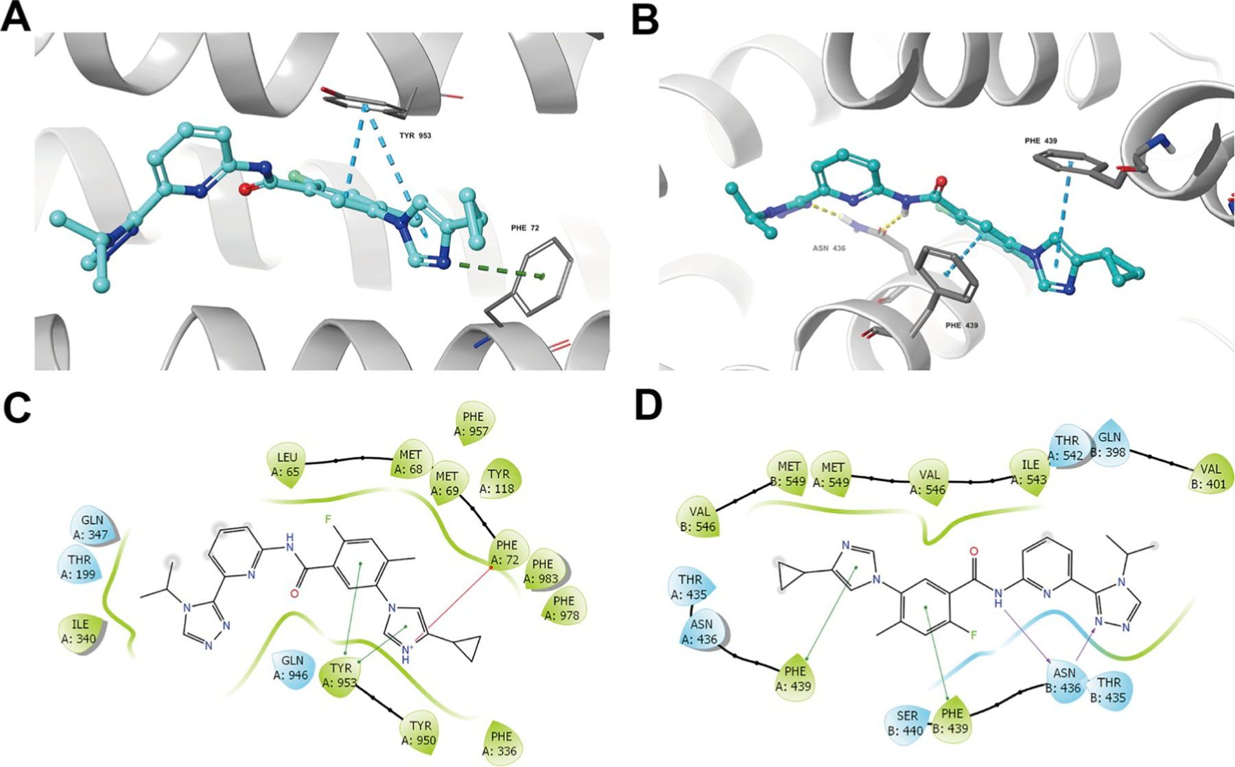
Fig. 8. In silico docking of selonsertib with homology model of human ABCB1 and human ABCG2.

(A) Docked position of selonsertib within the drug-binding site of ABCB1. (B) Docked position of selonsertib within the drug-binding site of human ABCG2. Selonsertib is shown as ball and stick mode with the atoms colored: carbon-cyan, hydrogen-white, nitrogen-blue, oxygen-red, fluorine-green, hydrogen-white. Important residues are shown as sticks with orange color. π-π stacking interactions are indicated with cyan dotted short line. Hydrogen bonds are shown by the yellow dotted line. The two-dimensional ligand-receptor interaction diagram of selonsertib and ABCB1 (C) and ABCG2 (D). The amino acids within 3 Å are shown as colored bubbles, cyan indicates polar residues, and green indicates hydrophobic residues. π-π stacking interactions are indicated with green short line. Hydrogen bonds are shown by the purple arrow. (For interpretation of the references to color in this figure legend, the reader is referred to the Web version of this article.)
