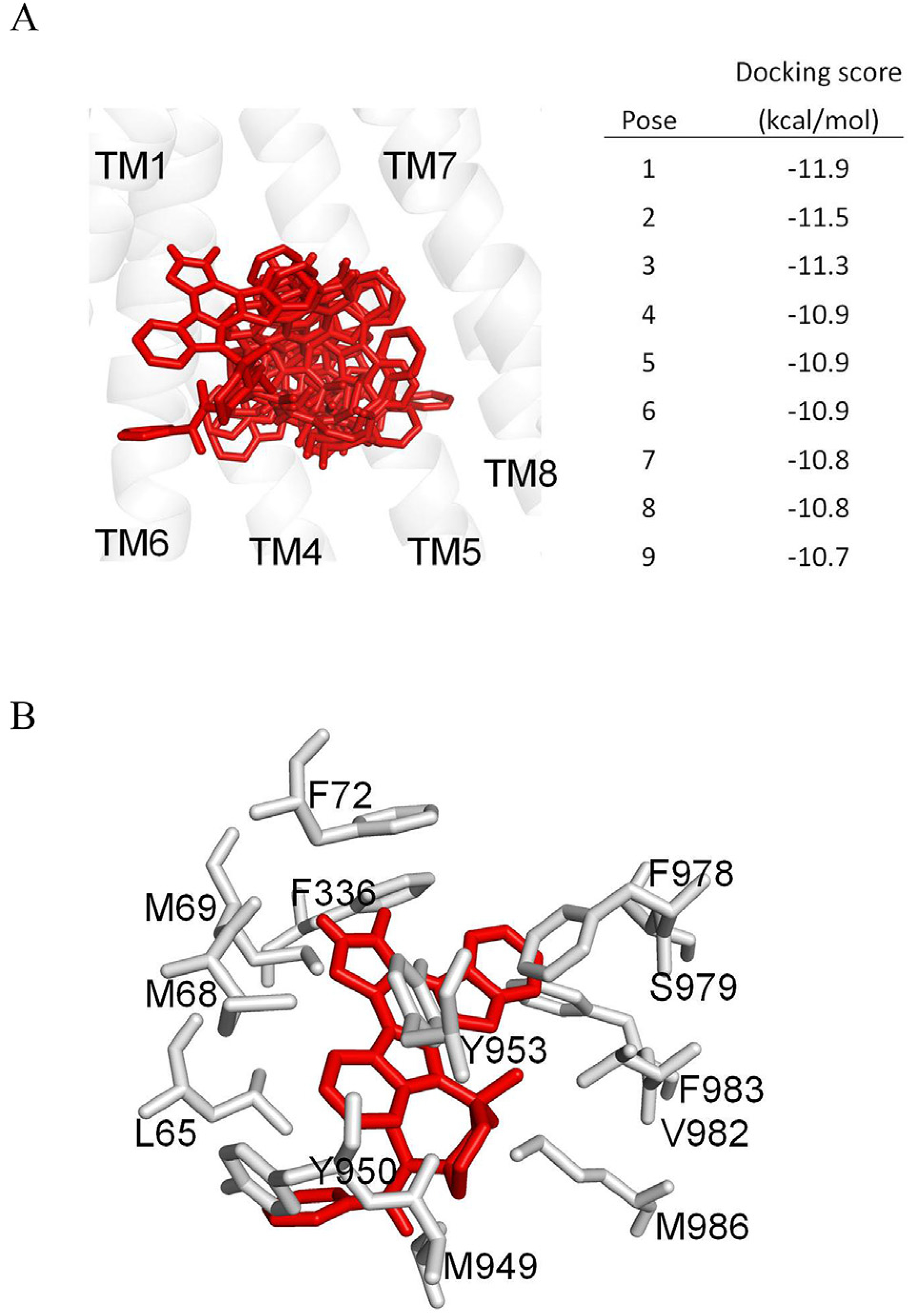
Fig. 6. Docking of midostaurin in the drug-binding pocket of ABCB1.

(A) The homology model of ABCB1, based on the PDB:5KPI mouse P-glycoprotein structure, was used for exhaustive docking using AutoDock Vina software. The receptor grid was centered at x = 20, y = 60 and z = 5 Å, and 33 residues in the binding-pocket were set as flexible. The box size was 50 × 50 × 50 Å and the exhaustiveness was set at 100. The first nine poses with the highest docking scores are shown in red sticks. For the purpose of clarity, only transmembrane helices 1 and 4 to 8 are presented in grey as a cartoon representation. The figure was prepared using PyMOL software. (B) The lowest docking score pose binding mode of midostaurin is presented in red sticks. Residues within 4 Å distance of midostaurin are shown in grey. (For interpretation of the references to color in this figure legend, the reader is referred to the Web version of this article.)
