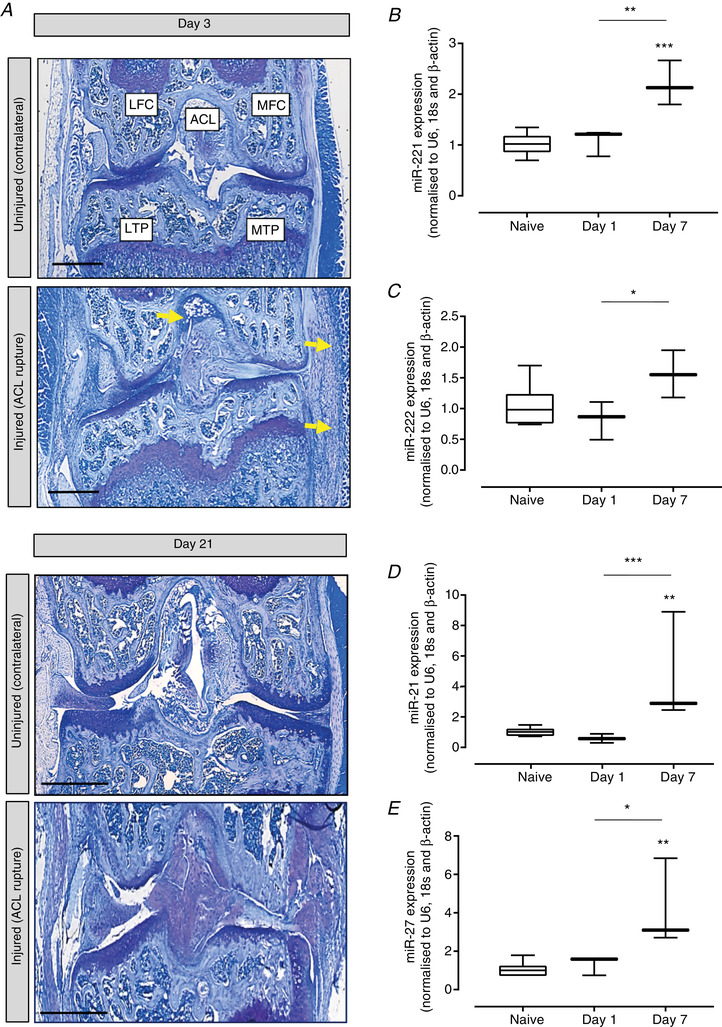
Figure 2. Validation of mechanically regulated miRNAs in a murine in vivo model of load‐induced joint degeneration.

A, toluidine blue staining of a representative mouse knee joint at days 3 and 21 after ACL rupture to induce joint instability/joint degeneration. MTP, medial tibial plateau; MFC, medial femoral condyle; LTP, tibial plateau; LFC, lateral femoral condyle; ACL, anterior cruciate ligament. Yellow indicates inflammatory cell infiltrate. Validation of differential expression of (B) miR‐221, (C) miR‐222, (D) miR‐21‐5p and (E) miR‐27‐5p in articular cartilage after normalization to the geometric mean of the reference genes U6, β‐actin and 18s and further normalization to the uninjured knee cartilage. Data are presented as box plots depicting the mean ± 95% CI (n = 3 animals per experimental time point). Statistical analysis was performed using one‐way ANOVA with Tukey's post hoc test. [Color figure can be viewed at wileyonlinelibrary.com]
