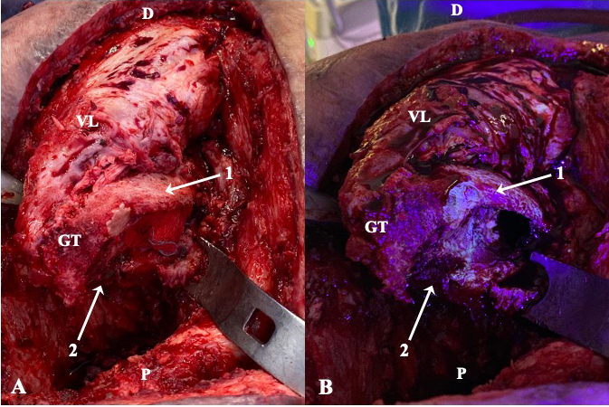
Figure 2.

Intraoperative clinical photographs showing the ventral aspect of the proximal femur of a patient operated through anterolateral approach, who had been receiving minocycline 100 mg every 12 h orally for 10 d (case 2). The surgical field prior (a) and after (b) the utilization of fluorescent light is compared. Under the black light, viable metaphyseal bone glowed greenish (1), whereas all that bone failing to fluoresce (2) was considered necrotic and thus removed. D: distal; P: proximal; GT: greater trochanter; VL: vastus lateralis.
