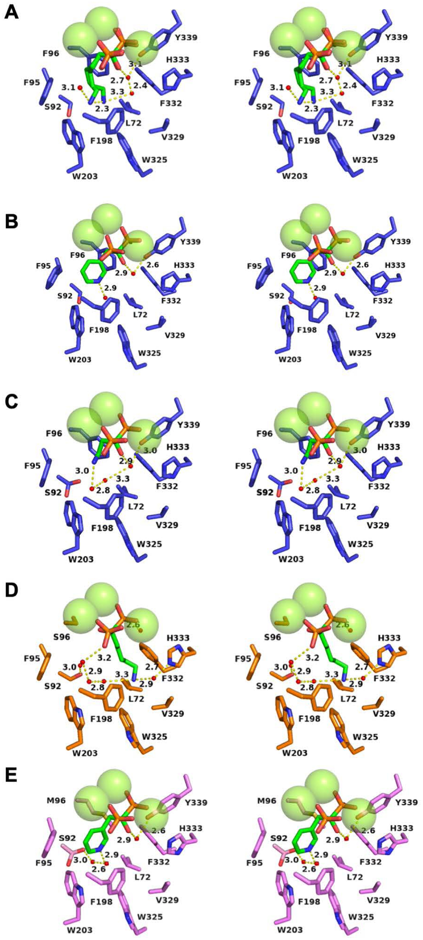
Figure 6:

Water molecules and solvent hydrogen bond networks in EIZS variants complexed with 3 Mg2+ ions and bisphosphonate inhibitors. Atoms are color-coded as follows: C = blue (wild-type EIZS), orange (F96S EIZS), violet (F96M EIZS), or green (inhibitors); N = blue; O = red; P = orange; water molecules = small red spheres; Mg2+ ions = large green spheres. Hydrogen bonds are indicated by yellow dashed lines and distances are reported in Å. (A) Wild-type EIZS–neridronate complex; (B) wild-type EIZS–risedronate complex; (C) wild-type EIZS–pamidronate complex (note that S92 adopts two alternative conformations); (D) F96S EIZS–neridronate complex; (E) F96M EIZS–risedronate complex (note that S92 adopts two alternative conformations).
