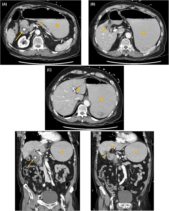
Figure 1 .

Axial (A–C) and coronal (D, E) computed tomography images showing gastric outlet obstruction (cross) due to a 3 cm hyperdense ectopic gallstone between the first and second parts of duodenum (arrow), with intrahepatic pneumobilia (diamond) and a decompressed gallbladder (arrow heads). This Rigler’s triad of findings was consistent with a cholecystoduodenal fistula and Bouveret syndrome.
