Figure 1.

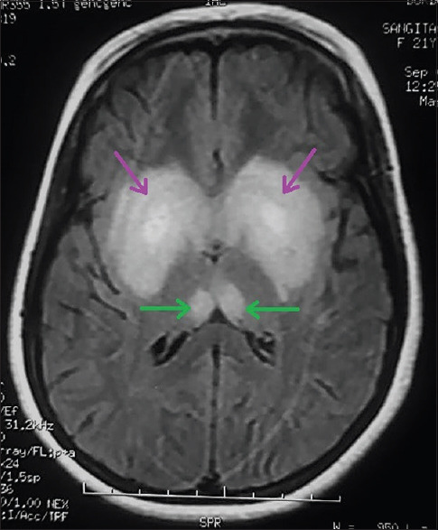
Cerebral MRI showing FLAIR hyperintensity of both dorsomedial thalami (green arrows) and both basal ganglia (purple arrows)

Cerebral MRI showing FLAIR hyperintensity of both dorsomedial thalami (green arrows) and both basal ganglia (purple arrows)