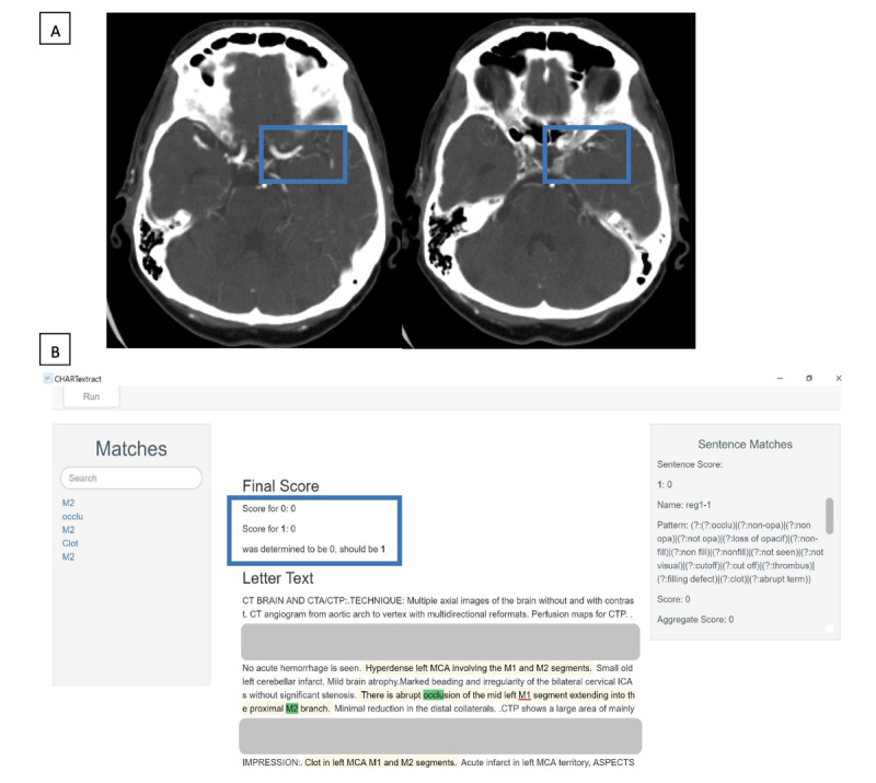
Figure 1.

Example 1 of a discrepancy between the chart abstractor and CHARTextract tool output. (A) Computed tomography angiography scan showing loss of opacification in the left middle cerebral artery, involving the left M1 segment and extending into the M2 segment. (B) CHARTextract tool output: the chart abstractor labeled that large vessel occlusion was present, but the CHARTextract tool determined this attribute to be absent. The rules were revised to reflect that occlusion involving the “M1 segment” should be considered a large vessel occlusion even if the terms “MCA” or “middle cerebral artery” were absent.
