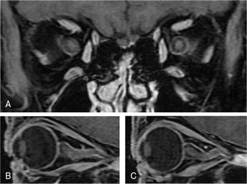
Figure 1.

Postcontrast T1-weighted fat-suppressed magnetic resonance imaging (MRI) of the orbits. (A) Coronal MRI of the orbits reveals bilateral (but left dominant) uniform enhancement of the optic nerve. (B) Sagittal MRI of the right orbit reveals a slightly ill-defined appearance of the optic nerve and slight enhancement of optic nerve sheaths. (C) Sagittal MRI of the left orbit reveals uniform enhancement along with optic nerve sheaths.
