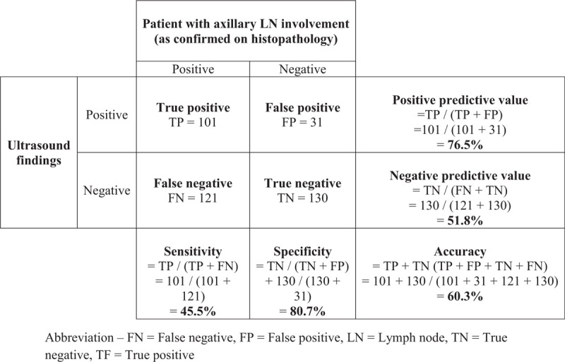
Figure 1.

Comparison of axillary lymph node status as assessed by axillary ultrasound and histopathology (n = 383). FN = False negative, FP = false positive, LN = lymph node, TF = true positive, TN = true negative.

Comparison of axillary lymph node status as assessed by axillary ultrasound and histopathology (n = 383). FN = False negative, FP = false positive, LN = lymph node, TF = true positive, TN = true negative.