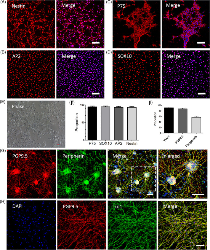
FIGURE 2.

Characterization of the identity and the differentiation capability of mouse premigratory neural crest stem cells (pNCSCs). A, The supposed pNCSCs at P2 are identified with immunofluorescence staining using the neural stem cells (NSCs) marker, Nestin (A‐left image), and the NCSCs markers P75 (B, left), AP2 (C, left), and SOX10 (D, left). These cells are counterstained with DAPI in the nuclei and showed merged images (A‐D, right image). The cultured cells at P2 are shown as the phase image (E). Most cells are immunoreactive to Nestin (93.2% ± 4.8%), P75 (94.5% ± 3.5%), AP2 (93.1% ± 5.0%), and SOX10 (94.6% ± 4.1%) (n = 3 independent experiments) as showed in the cartogram (F). G,H, Immunofluorescence staining of pNCSCs following cultured in differentiation medium II for 7 to 14 days. The differentiation into neurons is confirmed by the double immunostaining of pan‐neuronal markers PGP9.5 (red), peripheral neuronal marker peripherin (green, pseudo‐color), and the merged image of DAPI, PGP9.5, and peripherin staining. TuJ1 (green, pseudo‐color) and PGP9.5 staining with DAPI are also demonstrated (H). I, The proportion of positive cells in the TuJ1 staining (TuJ1, 91.93% ± 3.7%), PGP 9.5 (87.3% ± 5.4%), and peripherin (57.3% ± 7.0%). Scale bars = 100 μm
