
FIGURE 4.
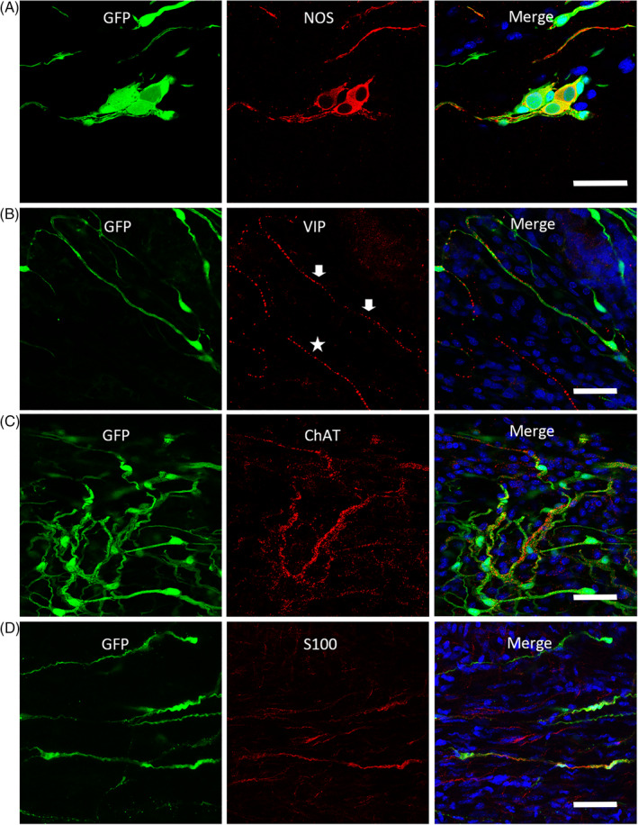
EGFP+ cells differentiated into glia cells expressing S100β, and neurons expressing nitric oxide synthase (nNOS), Vasoactive Intestinal Peptide (VIP), and choline acetyltransferase (ChAT) at 2 months after transplantation. A‐C, The colocalization of grafted EGFP+ cells with the immunofluorescence staining of enteric neurochemical markers nNOS, VIP, and ChAT. B, The arrows indicate the VIP staining with EGFP fluorescence, and the asterisks show VIP expression of endogenous neurons. D The fluorescence of some EGFP+ cells is colocalized with the immunostaining of glia cell marker, S100β. Sections of the mouse colon are counterstained in blue with DAPI. Scale bars = 50 μm