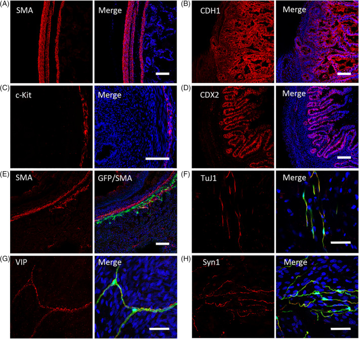
FIGURE 6.

Immunofluorescent staining of tissue‐engineered intestine (TEI) and TEI + premigratory neural crest stem cells (pNCSCs). A, The positive immunostaining of smooth‐muscle marker (SMA) is found in the locations of myenteric and submucosal layers in the TEI sample. B, The positive immunostaining of the epithelium marker CDH1 is showed in TEI. C, Immunostaining of c‐Kit, a marker of interstitial cells of Cajal (ICCs). D, Immunostaining of CDX2, a marker expressed in the nuclei of epithelial cells throughout the intestine. E, The distribution of EGFP+ cells in the muscular layer (expressing SMA) of TEI. F‐H, The positive immunofluorescent staining of pan‐neural marker TuJ1 (F) and marker of enteric neuron subtype VIP (G) are available as showed by co‐expression with EGFP in the muscular layers of pNCSCs‐TEI, Synapsin‐1 was also found coexpressed with EGFP+ cells in the myenteric layers (H). Scale bars (A‐E) = 100 μm and (F‐H) 50 μm
