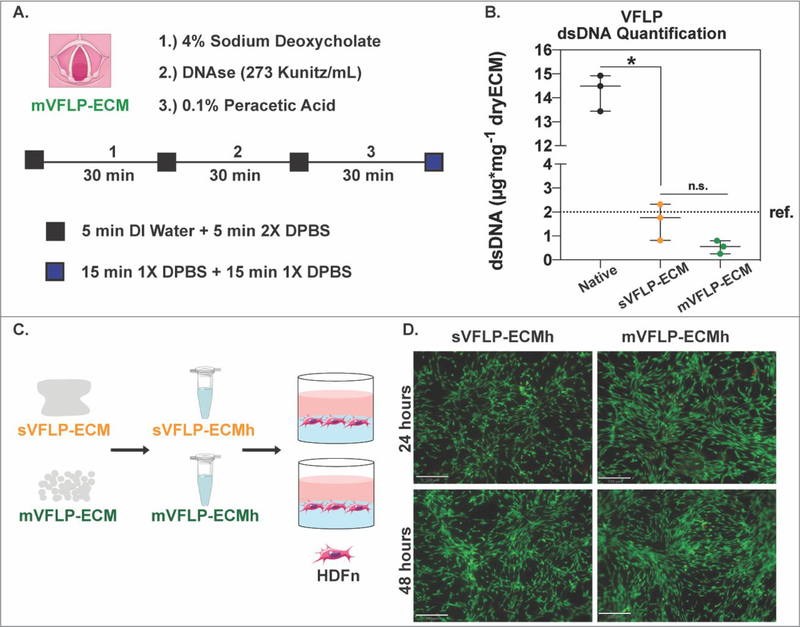
Figure 8. Vocal fold lamina propria decellularization.
A.) Schematic showing the protocol for the automated decellularization of porcine vocal fold lamina propria (mVFLP-ECM). B.) Box and Whisker plot showing dsDNA quantification per mg of dry tissue for native and decellularized sVFLP-ECM and mVFLP-ECM. Approx. 40 VFLPs were combined from 20 animals and the decellularization was performed three independent times (n=3). * = P<0.05. n.s. = not significant (P>0.05). ref. ~2μg/mg (for Urinary Bladder Matrix–UBM). C.) ECM scaffolds were enzymatically digested, self-assembled into ECM hydrogels, and seeded with HDFn for cytocompatibility studies. D.) LIVE/DEAD imaging of HDFn cells cultured on sVFLP-ECM, and mVFLP-ECM. Calcein-AM staining (Green) shows live cells while Ethidium Homodimer-1 staining (Red) shows dead cells (Scale bar = 330μm).

