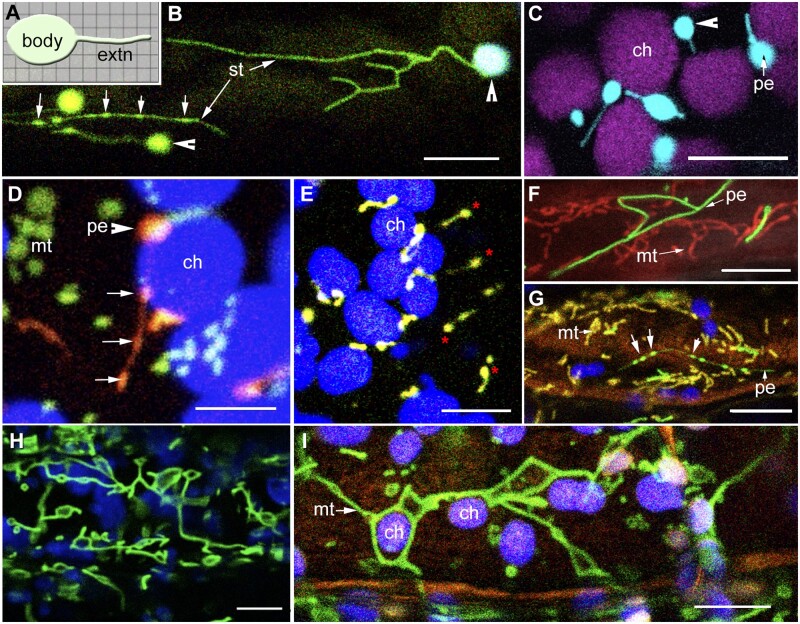
Figure 1.
Characteristic pleomorphy of organelle extensions and tubular organelles in plant cells. (A) Diagrammatic depiction of an organelle and its tubular extension (extn). The cytoplasm is represented by the grid in the background. Key features involve an independent organelle body from which tubules extend and retract. Cytoplasmic outreach of the organelle increases during tubule extension. (B) Snapshot showing shape diversity displayed by stroma filled tubules (st) extended from plastid bodies (arrowheads) including the presence of dilated regions (arrows) in two stromules (Supplemental Movie S1). (C) Tubular peroxules extended from the peroxisome (pe) body (arrowhead) in the cortical region lying above chloroplasts (ch; Supplemental Movie S2). (D) Snapshot from a time-lapse series showing punctate mitochondria (green; mt), peroxisomes (orange; pe), and chloroplasts (blue; ch). Dilated regions (arrows) appear in an extending peroxule (Supplemental Movie S3). (E) Peroxules (red asterisks) are formed in response to increased ROS in a cell and may be extended by peroxisomes that are not appressed to chloroplasts. (F) Snapshot of abnormally elongated peroxisomes (pe) and mitochondria (mt) resulting from lack of efficient fission in the Arabidopsis apm1/drp3a mutant. (G) Similar to their formation in different organelle extensions, dilations also appear in tubular organelles (large arrows) such as the elongated peroxisomes (green, pe) and mitochondria (yellow, mt) in the Arabidopsis apm1/drp3a mutant. (H) Under hypoxia mitochondria (green) transiently stop dividing and instead expand into assorted shapes. (I) Expanded mitochondria (green, mt) are able to fuse and frequently encompass large organelles like chloroplasts (ch). Scale bars in µm: D = 5; all others =10.

